Teacher Portal:
Microscopes and Magnification
Investigation 3
Microscopes and Magnification: Investigation 3
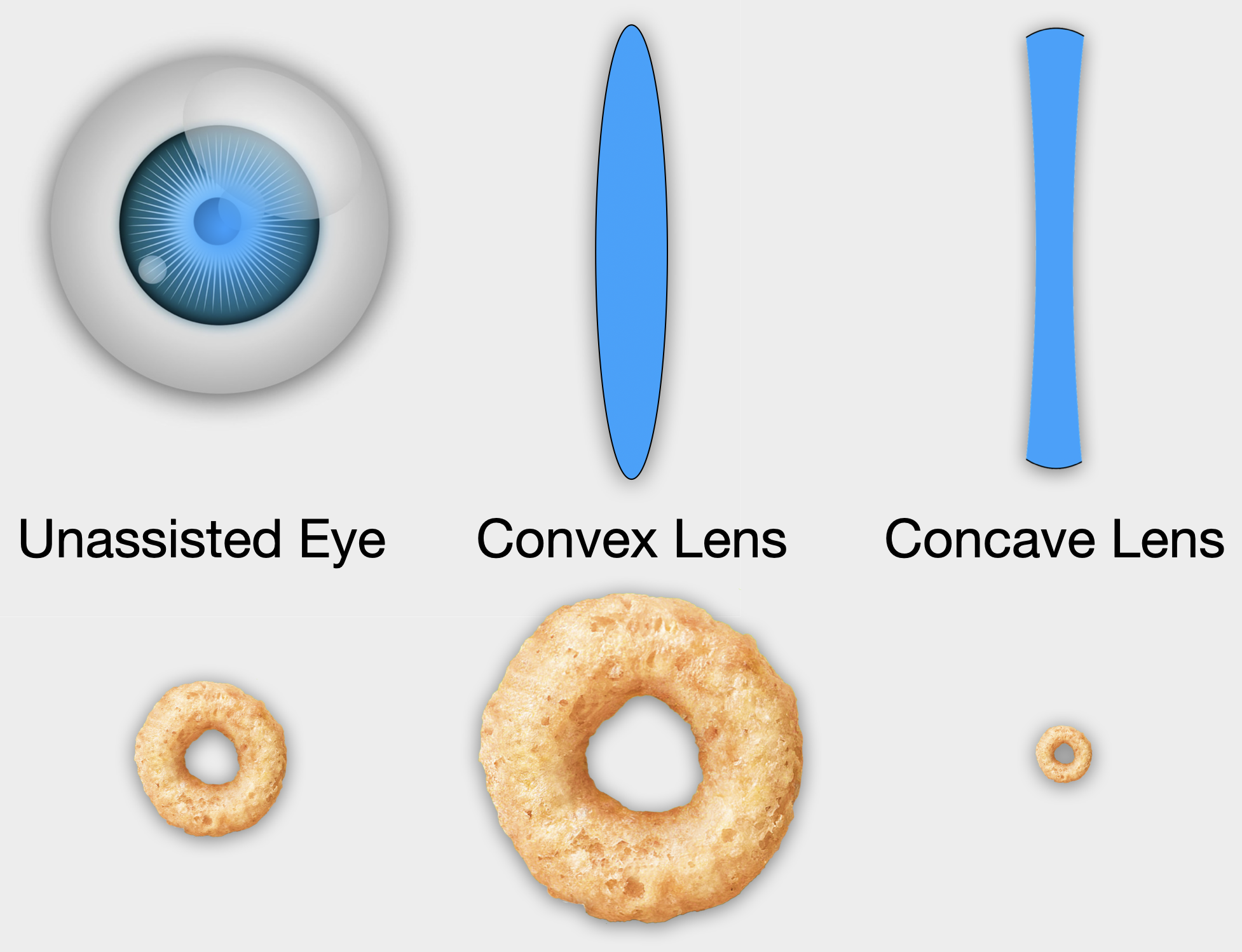 In Investigations One and Two, you explored how lenses refract light and change the apparent size of an object by comparing the images of objects viewed with the naked eye or with the assistance of the hand lens. You also investigated how the refraction of light by a convex and a concave lens results in the magnification or the reduction of the size of an object’s image. In this Investigation, you will learn about the history and operation of the compound microscope, a sophisticated scientific tool that magnifies the image of objects too small to see with the naked eye. Through the use of more than one convex lens, the microscope possesses a higher total magnification than a hand lens.
Scientists use the microscope in microscopy, the science of viewing exceedingly small or microscopic objects. Scientists use the microscope to assist in their research of plants, animals, bacteria, and viruses. Medical professionals use the microscope to diagnose the cause of infections and to determine the presence of abnormal cells obtained from an ill patient.
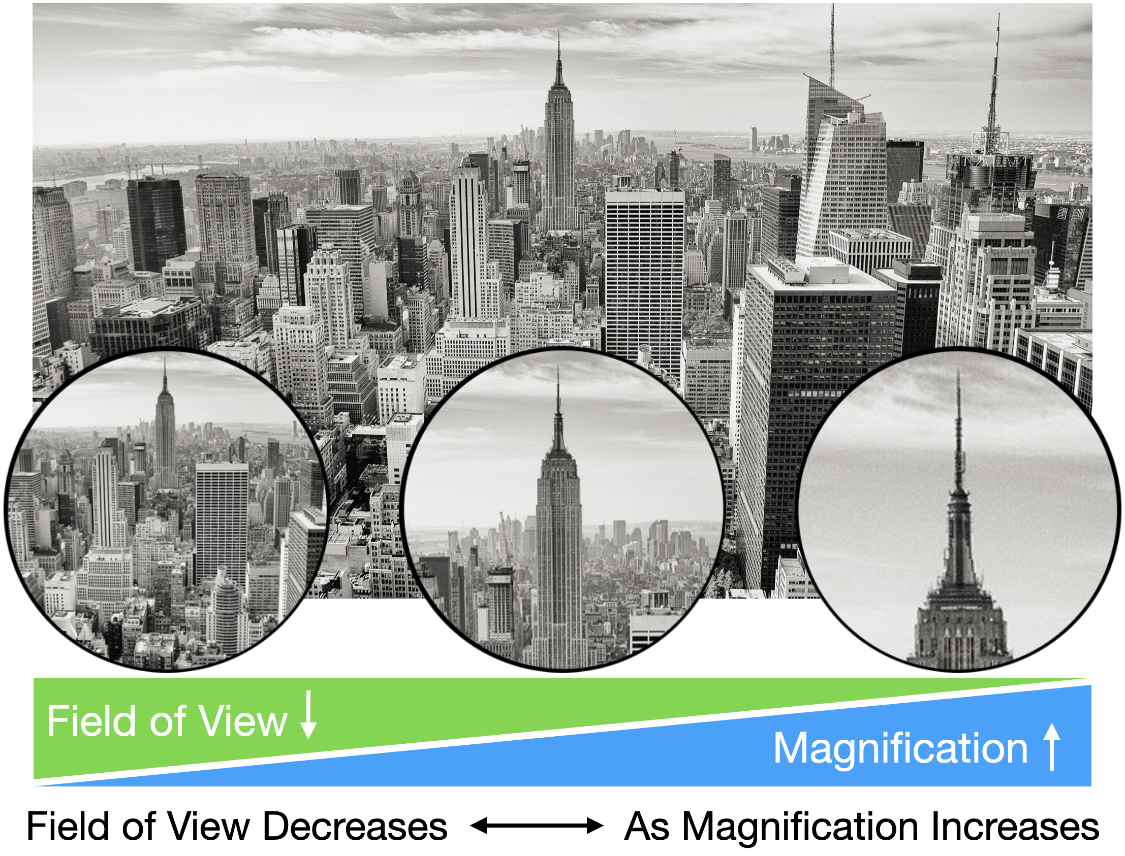
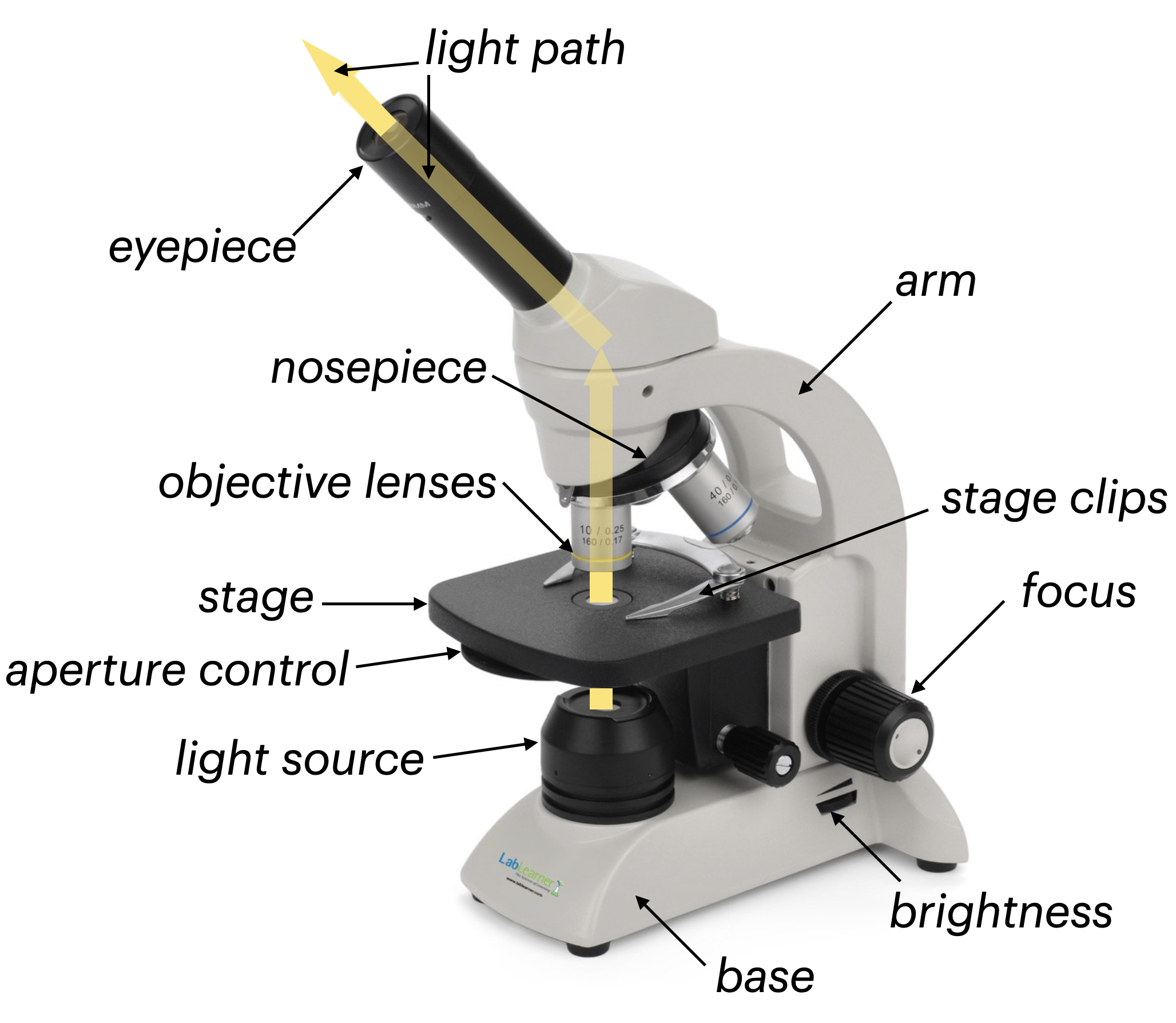 A slide is a small piece of glass used to hold a specimen, a sample of the object to be viewed. An important consideration when viewing a specimen using a microscope is that the specimen must be thin enough to allow light to pass through it, otherwise, nothing can be seen. A slide is secured on the stage of the microscope, which is located below its objectives, each of which contains convex lenses. Each of the three objectives has a different power of magnification. The powers magnification of most compound microscopes is 4x, 10x, and 40x. The image of a specimen is viewed through the eyepiece, which also possesses a convex lens with a 10x power of magnification. The total power of magnification for each objective is the product of the magnification of the objective and the eyepiece or 40x, 100x, and 400x. The portion of the specimen visible through the eyepiece, also called the field of view, changes when a different objective is used, so more or less of the complete specimen will be visible.
Each time a specimen is viewed through the microscope, you will learn to follow a specific procedure to avoid damaging the microscope or the slide. You will identify the parts of the microscope and gain experience using the microscope so that you are able to compare the capabilities of the microscope to other magnification tools.
Microscopes and Magnification : Investigation 3 - Mathematics Concepts
PreLab
• problem-solving
Lab
• parts/whole
• sequential order
• whole numbers
• Venn Diagram
Postlab
• multiplication
• Venn Diagram
• Data analysis
• Problem solving
• Greater than/less than/equal to
Microscopes and Magnification: Investigation 3 - Procedural Tools

Microscopes and Magnification:
Investigation 3 Quiz

