Teacher Portal:
The Human Body
Investigation 4
The Human Body: Investigation 4
Investigation One introduced students to the various roles of the nervous system in controlling the functions of the remainder of the body’s physiological systems. Students conducted experiments to study the sensory nervous system, the autonomic nervous system, memory, and thought processes. In Investigation Two, students continued their exploration of the human body by examining the skeletal and muscular systems by testing a model of a joint and muscle, and exploring a model of a human torso. Investigation Three introduced students to the respiratory system through experiments involving respiratory rate and lung capacity. Investigation Four introduces students to the circulatory system.
 If the nervous system can be considered the body’s command and control center, then the circulatory system can be considered the supply system. The circulatory system consists of the heart, arteries, veins, capillaries, and blood.
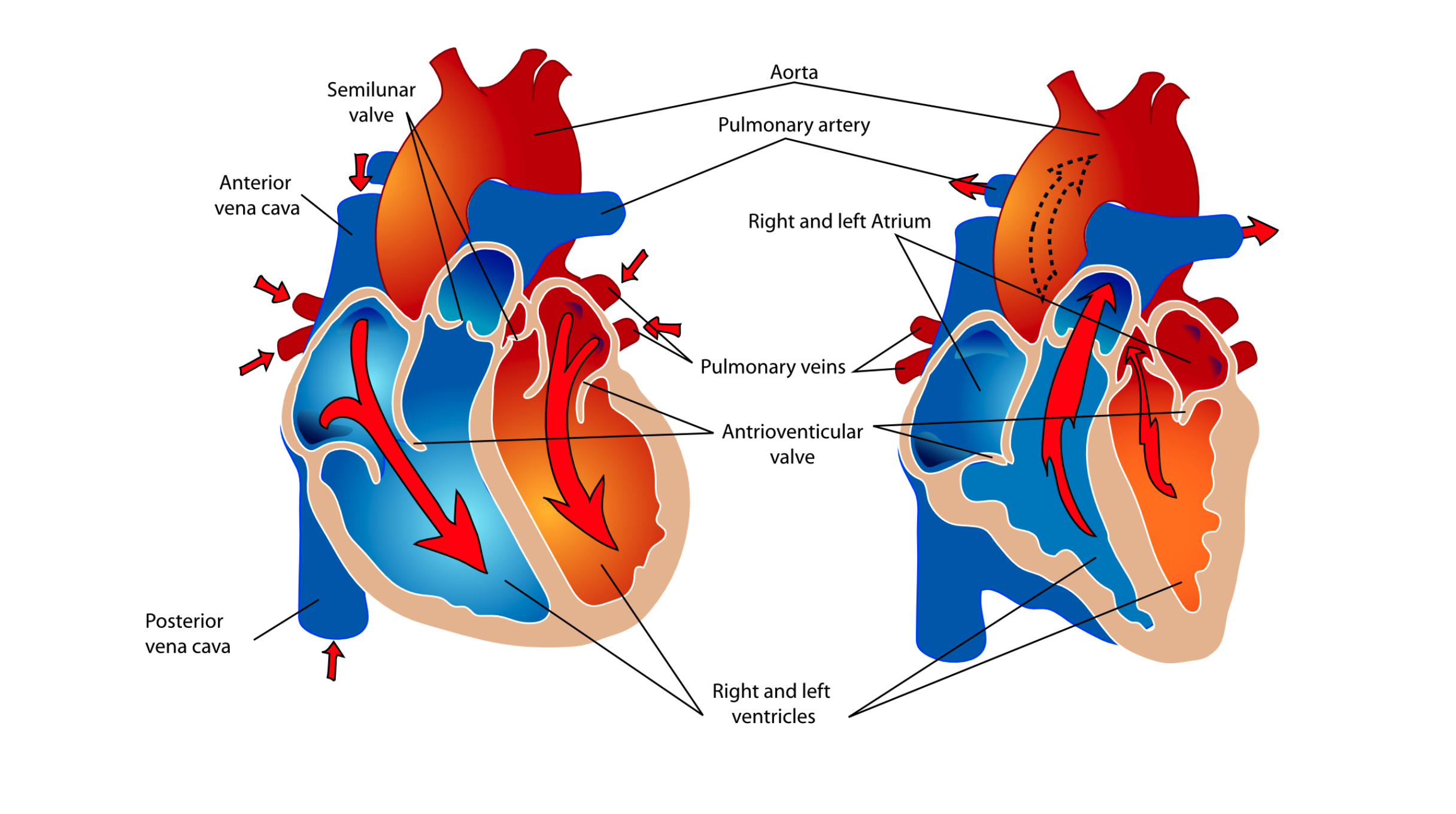
The heart is the distribution center of the body. It has four chambers that alternately collect and redistribute blood. The top two chambers are called atria (singular atrium). De-oxygenated blood enters the heart through the right atrium. The right atrium then contracts and moves the blood into the right ventricle. Contraction of the right ventricle sends blood to the lungs via the pulmonary artery where it releases carbon dioxide and picks up oxygen. Blood then returns to the left atrium through the pulmonary vein. The left atrium pumps oxygenated blood into the left ventricle, which contracts and sends blood out into the body. If you were to lay out all of an adult’s arteries, capillaries, and veins, end-to-end, they would stretch about 100,000 kilometers. That would circle around Earth more than twice!
To summarize:
- Blood that has circulated throughout the circulatory system returns to the heart and enters the right atrium.
- The right atrium contracts and forces the blood through the tricuspid valve into the right ventricle.
- The right ventricle contracts and forces the blood to the lungs through the pulmonary valve.
- Blood is oxygenated in the lung and returns to the heart through the mitral valve and into the left atrium.
- The left atrium contracts and forces the oxygenated blood into the left ventricle.
- The left ventricle contracts and forces the oxygenated blood out of the heart through the aortic valve and from there, throughout the entire body.
The force of the contracting left ventricle must be very strong as blood leaving it must course through the entire circulatory system and then return to the heart. The outer wall of the left ventricle is typically thicker and stronger than the left ventricle wall, which only pumps blood to the lungs. You can follow the flow of blood through the heart by playing the video below. Play it a few times and you’ll begin to see how blood circulates through the heart.
The blood vessels serve as the body’s transport system. Blood exits the left ventricle of the heart into the aorta. Arteries are blood vessels that carry blood away from the heart. The aorta is the main artery of the body trunk. Blood is distributed to the body from the aorta by a series of major arteries. The carotid artery carries blood to the brain. Brachial arteries serve the arms, and femoral arteries carry blood into the legs. Major organs also have arteries branching off the aorta, such as the renal and hepatic arteries, which serve the kidneys and liver respectively.
Blood passes through a series of smaller arteries to tiny blood vessels called capillaries which deliver blood directly to the various body tissues. In most cases, oxygen moves out of the capillaries into the tissues and carbon dioxide moves into the blood from the tissues. In the lungs, however, this process is reversed at the alveoli. Here, oxygen moves into the blood while carbon dioxide moves out of the blood.
In areas of the body except the lungs, deoxygenated blood moves from capillaries into the veins. Veins are responsible for carrying blood back to the heart. Blood moves from the smallest veins into larger and larger veins to be collected into one of the two main veins serving the trunk of the body. These main veins are called the venae cavae (singular vena cava), which join together at the top of the right atrium.
Blood
The body uses blood to transport nutrients and oxygen to the cells, and waste products, such as carbon dioxide, away from the cells. Because body cells, tissues, and organs need oxygen in order to function, the rate at which oxygen-containing blood can be delivered  to all parts of the body can be regulated by the circulatory system. This occurs through changes in heart rate or the number of times the heartbeats per minute. Increases in heart rate increase the amount of blood and oxygen that each organ and body part receives. Decreases in heart rate decrease the amount of blood and oxygen that each organ or body part receives.
When the body is at rest, the heart rate is lower than during times of exercise or exertion when the muscles and other organs of the body require more oxygen to function. The average resting heart rate for adults is 60–80 beats per minute and increases to 80–100 beats per minute with moderate exercise. The heart rate of children tends to be higher than that of adults. The typical resting heart rate for elementary school-age children is 80–100 beats per minute and increases to 100–120 beats per minute during exercise.
The blood that is delivered to the body’s organs during rest or exercise appears to be a fluid but is technically a tissue because it consists primarily of cells. Two major types of cells are found in blood. Red blood cells (labeled RBC in video shown here) transport oxygen and carbon dioxide.

Mature red blood cells are unique compared to other cells in the body because they do not have a nucleus. Red blood cells are manufactured in the bone marrow, which is found in long bones as well as ribs and vertebrae. Red blood cells have a life span of about four months, and therefore are continually being replaced. Red blood cells contain a special protein called hemoglobin which binds oxygen and carbon dioxide for transport. White blood cells (labeled WBC in the video shown here) are part of the body’s immune system. The immune system is the body’s self-defense mechanism. White blood cells are carried to sites where disease organisms manage to enter the body, where they attack the disease-causing cells.
 A third component of blood is the platelet (labeled P in the video above). The platelet acts as “glue” when an injury to a blood vessel occurs. Platelets have a sticky surface that catches on the rough edge of a damaged blood vessel wall. Other platelets stick to the first platelet, creating a plug to prevent blood loss. The liquid portion of the blood is called the plasma. The plasma contains clotting proteins, nutrient transporters, nutrients, and water. It also contains products of nutrient metabolism which need to be removed from the body. These waste products are removed when blood passes through the kidneys.
In Investigation Four, students will explore the circulatory system by using stethoscopes to listen to their hearts beating. Students will use models to help visualize how the contractions of the heart are correlated to the sounds they hear through the stethoscope and will investigate the effect of exercise on heart rate and oxygen delivery to the body.
The Human Body: Investigation 4 - Mathematics Concepts
Prelab
- counting whole numbers
- grouping
- parts/whole
Lab
- counting whole numbers
- parts/whole
- sequential order
- grouping
- estimate/verify predictions/measurements
- time in seconds/minutes
- data table
- rate
Postlab
- rate
- counting whole numbers
- time in seconds/minutes
- interpreting data table
- grouping
- place value (ones, tens, hundreds)
- addition
- comparison
- parts/whole
- problem-solving
The Human Body: Investigation 4 - Procedural Tools

The Human Body: Investigation 4 - Cognitive Tools
The Human Body:
Investigation 4 Quiz

