Teacher Portal:
The Human Body
Investigation 4 – PostLab
ASK WHY

The human body is organized into systems that are made up of many parts and that these systems each perform both individual and complementary functions that occur at the same time in the body.
BRANCH OUT
Biomedical research scientists study the function of normal and diseased body systems. They study the human body at the systems level, the cell level, and the molecular level. Great medical advances have come from the understanding of the human body discovered through biomedical research.
PRINT IT
Use your browser to download a printable PDF as help during the slide presentation and to make additional notes. In your browser, go to File > Print and then choose to save as PDF.
NAVIGATE IT
Once the slide presentation is launched
- use your left and right arrows to advance or go back in the slide presentation, and
- hover your mouse over the left edge of the presentation to get a view of the thumbnails for all the slides so that you can quickly move anywhere in the presentation.
- Click HERE to launch the slide presentation for the CELL.
SHARE IT

SLIDE BODY-4post-1
In this PostLab session, we will review the circulatory system and the results of Trials 1 through 3 from the Lab for Investigation 4.
______________________________________________
SLIDE BODY-4post-2
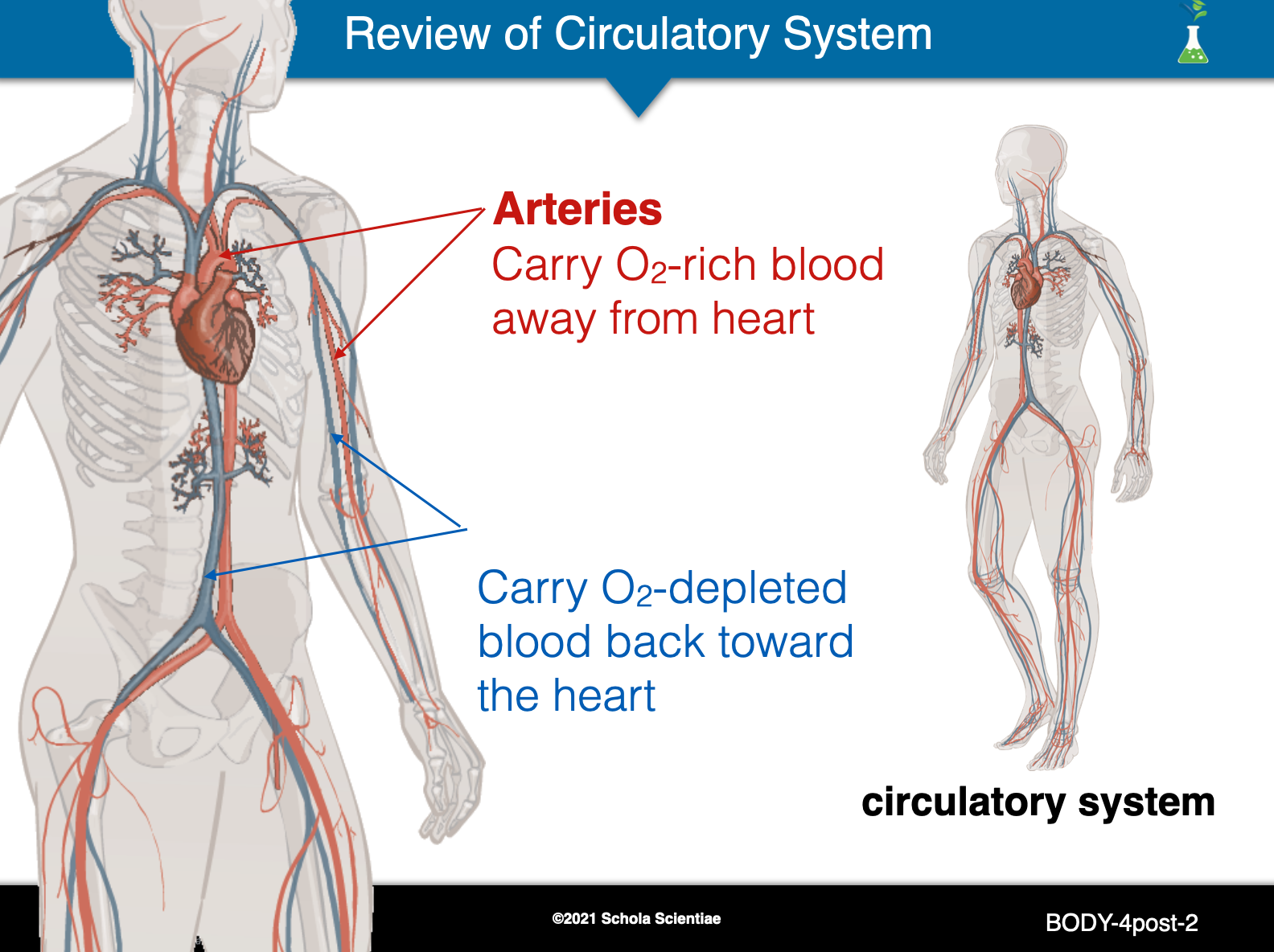
- Review the three main components of the circulatory system: heart, veins, and arteries.
- Ask students to look at the diagram of the circulatory system in problem1 of their Scientist Data Record. Using this diagram, students should label the heart, veins, and arteries.
- Use the human torso and the flip chart to aid students in labeling the diagram. Ask a student volunteer to point to the different parts on the torso and the flip chart as students are labeling their diagram. As the pieces of the human torso are removed, the red arteries and blue veins can be seen throughout the organs. This should be brought to the attention of the students.
- Explain to students that you are going to review the path that blood takes as it moves through this system specifically from the heart to the hand. As you discuss this pathway, point out the different parts on the human torso and/or flip chart. Encourage students to trace the pathway on the diagram in problem 1 with their fingers as it is discussed.
- Veins pump blood into the left side of the heart. This blood is full of oxygen.
- The blood flows away from the heart through the arteries.
- The oxygen-rich blood makes its way through the arteries of the arm and ends up in the hand.
- Once the blood reaches the hand, the oxygen is dropped off to the muscles so that the hand can move.
- The blood leaves the hand and starts to make its way back to the heart through the veins of the arm. This blood has very little oxygen.
- The blood empties from the veins into the right side of the heart.
- The blood flows from the right side of the heart into the arteries of the lungs.
- The blood picks up oxygen in the lungs and returns to the left side of the heart through the veins of the lungs. The entire process begins again.
______________________________________________
SLIDE BODY-4post-3
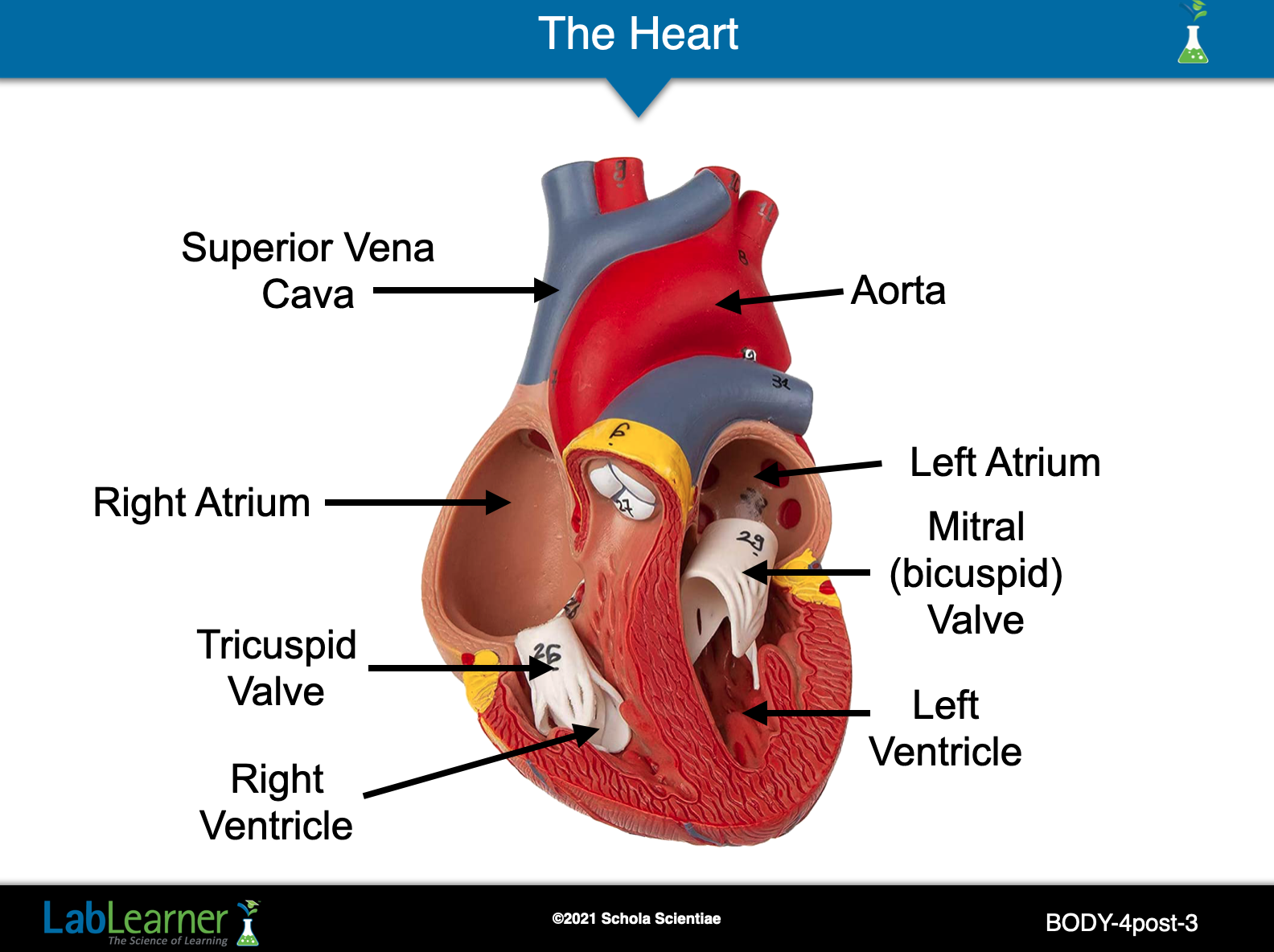
Ask students: Which part, or organ, of the circulatory system is responsible for pumping blood? Students should indicate that the heart is responsible for pumping blood through the veins and arteries.
Tell students that they are going to take a closer look at the heart. Turn the flip chart to the heart diagram. Remove the heart from the human torso and pull it apart (see above).
Ask students: What does your heart look like on the inside? Is it solid? Is it hollow?
Student answers may vary. Encourage students to realize that the heart is hollow and made up of four compartments or chambers. Two chambers are on the right and two chambers are on the left. Blood is pumped through these chambers as it is leaving the heart and returning from the body.
Note: The interior of the human torso heart also shows the two heart valves: the tricuspid valve and mitral (bicuspid) valve. The valves permit the one-way flow of blood between each atrium and ventricle by preventing the blood in the ventricles from flowing back into the atria. It is optional to share this information with students.
______________________________________________
SLIDE BODY-4post-4
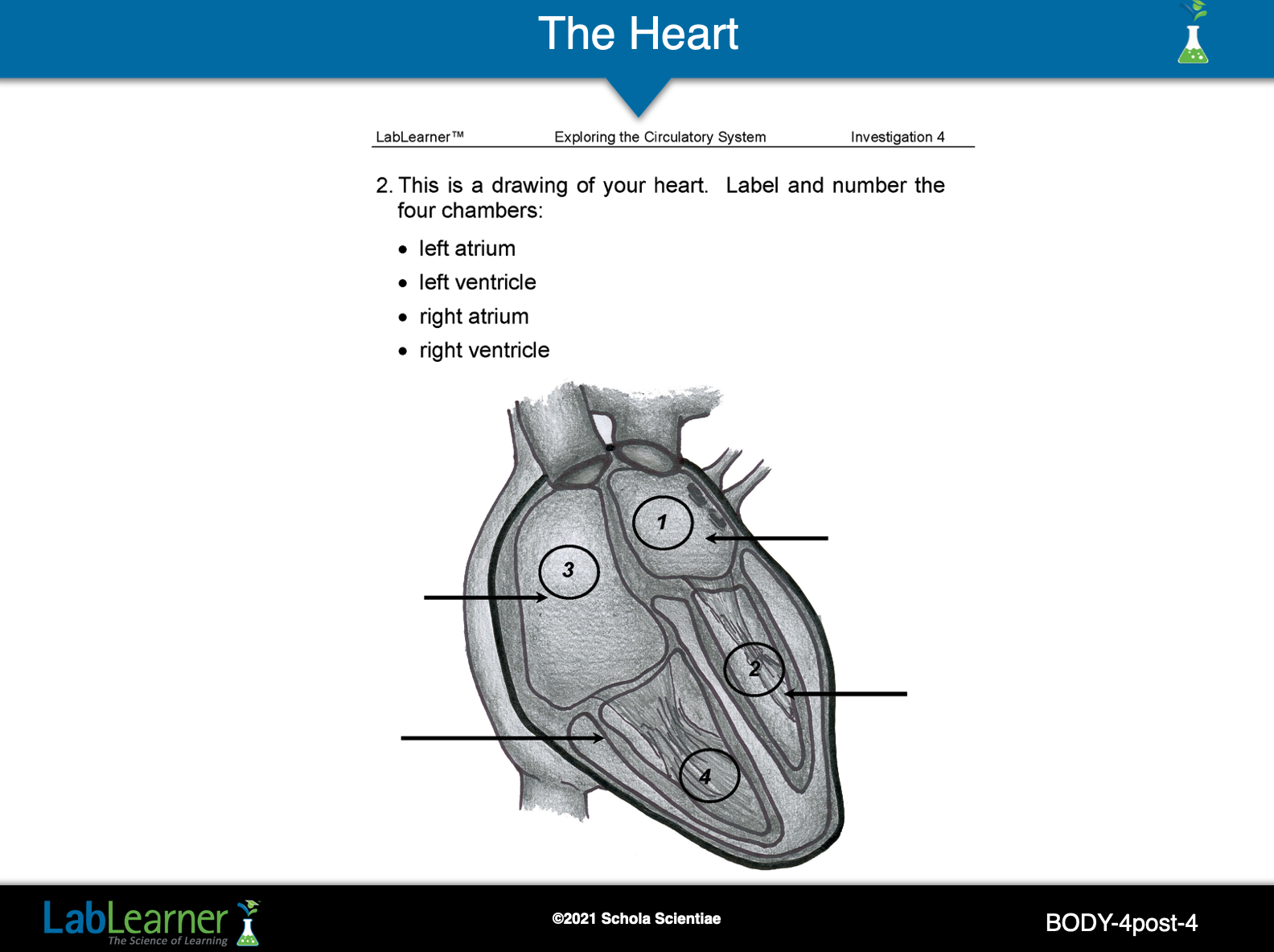
Tell students to find the picture of the heart in Problem 2 of their Scientist Data Record. As you point out the different chambers of the heart (left atrium, right atrium, left ventricle, right ventricle), encourage students to label them using the word bank at the top of the page.
Remind students that they are looking at a mirror image of the heart. The right side of the heart is located on the left side of the paper and the left side of the heart is located on the right side of the paper. Students should also number the four chambers by placing numbers one through four in a circle at the center of each chamber.
______________________________________________
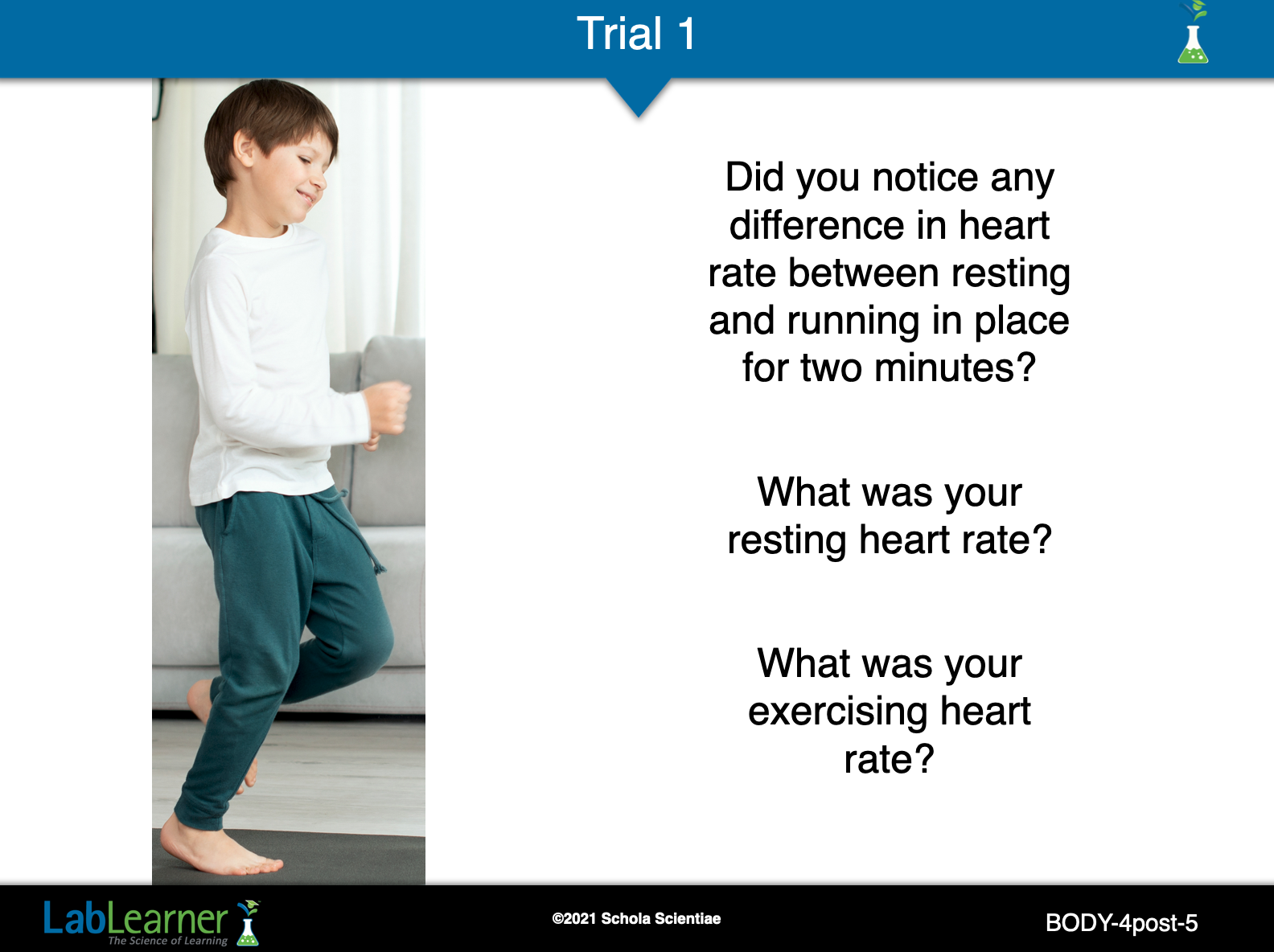
SLIDE BODY-4post-5
The heart pumps blood to all the parts of the body. It pumps oxygen-containing blood from the lungs to every cell in the body.
Ask students if they were surprised that their heart rate increased with exercise. Student answers will vary but most will not be surprised that exercise increases heart rate.
Ask students why they think the heart increases its rate during exercise. Student answers will vary, however, the main point is that when our muscles do work (exercise) they produce more waste products like carbon dioxide (CO2) and require more oxygen (O2).
______________________________________________
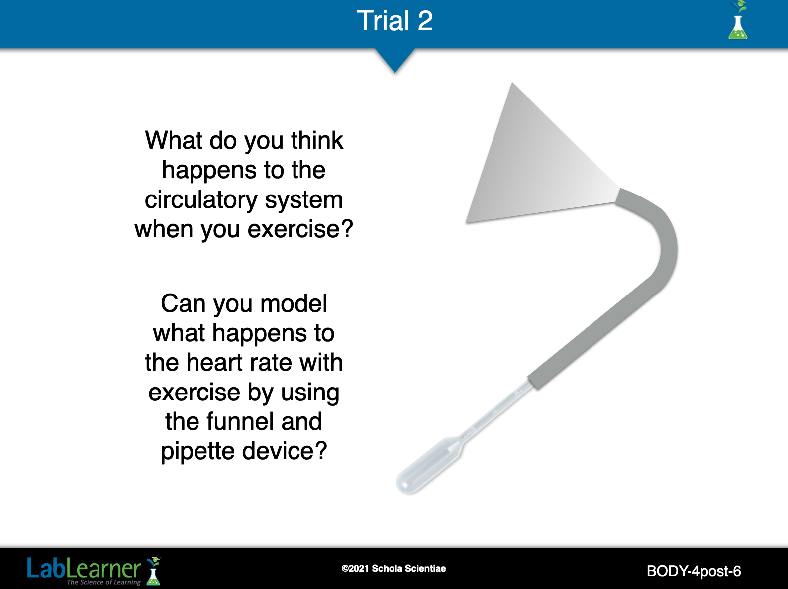
SLIDE BODY-4post-6
Use this slide to discuss what students did, saw, and heard in Trial 2.
______________________________________________
SLIDE BODY-4post-7
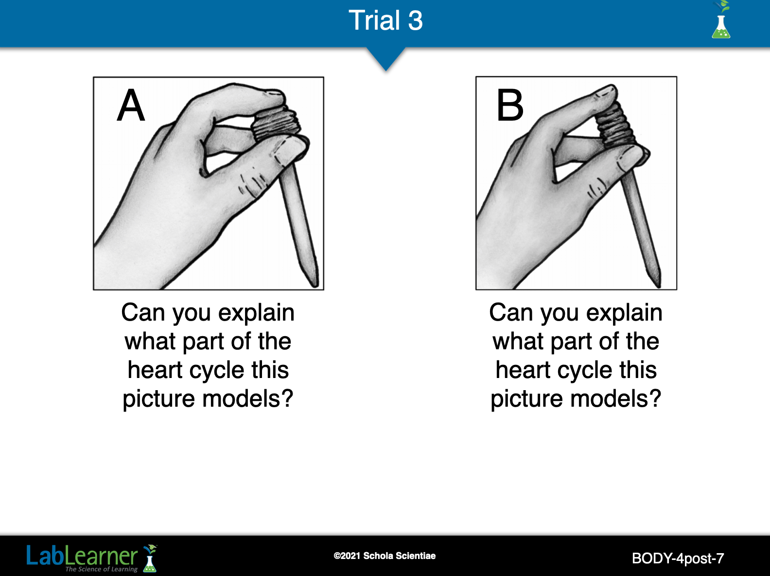
Use this slide to discuss what students did, saw, and heard in Trial 3.
Ask students to discuss the model of the heart using the plastic pipette:
In which example does the heart fill with blood? The model in B sucks blood into the heart.
In which example does the heart contract and relax? The model in B represents the heart in a relaxed state. The model in A shows the contraction of heart muscles that forces blood out of the heart.
Referring to the “Lub-Dub” sound used to describe the heart, which model (A or B) is the state associated with the Lub sound? Which state is associated with the Dub sound? Model A represents the “Lub” sound and model B represents the “Dub” sound.
______________________________________________
SLIDE BODY-4post-8
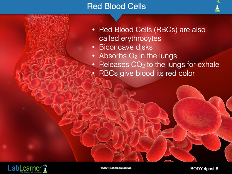
There are a number of different types of blood cells. White blood cells (leukocytes) are important for fighting infections. Platelets circulate in the blood and are important in forming blood clots.

In terms of our discussion of the blood carrying oxygen throughout the body however, the most abundant blood cell is the red blood cell, also called an erythrocyte. The video above illustrates red blood cells rushing through a blood vessel.
______________________________________________
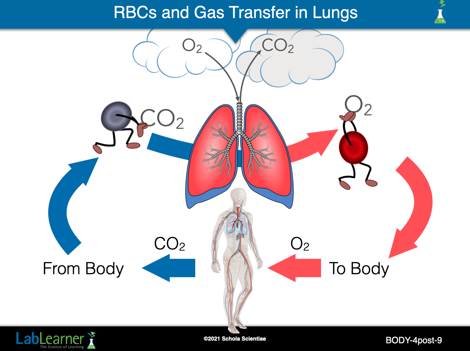
SLIDE BODY-4post-9
RBCs carrying CO2 waste from the body’s cells enter the lungs after circulating throughout the body. In the lung, the CO2 is released by the RBCs and is exhaled by the lung. Next, the RBC binds to an oxygen molecule (O2) breathed in through inhalation by the lungs.
This process is repeated by the RBCs over and over until they are worn out and replaced. Normal RBCs last about 121 days before they are replaced.
______________________________________________
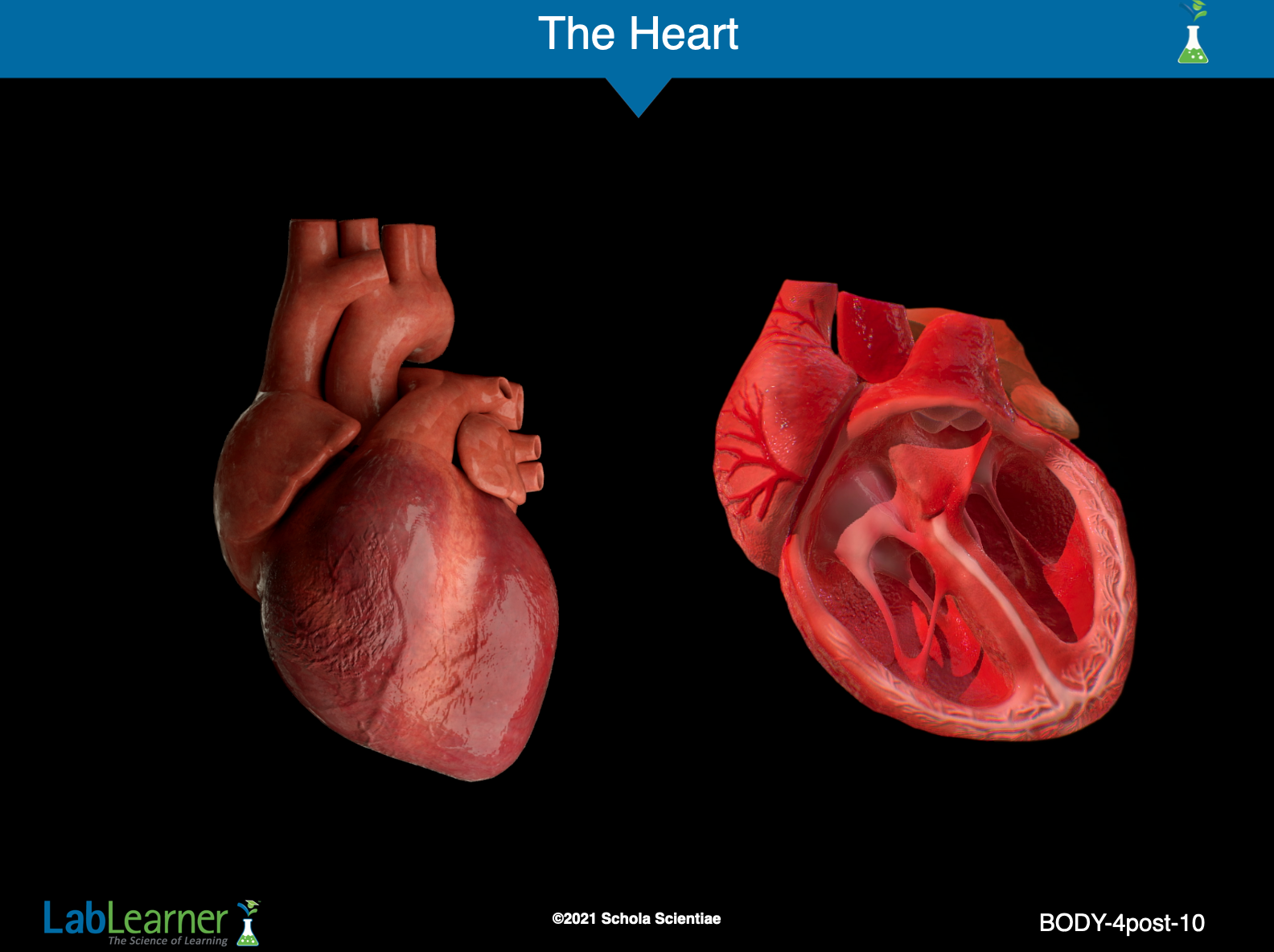
SLIDE BODY-4post-10
While this slide is up, ask the questions that follow to prompt students to Recall and share their previous knowledge on this topic.
1. Ask students: What do you know about the heart? Student answers may vary. Allow time for students to share their ideas.
2. Tell students that the heart is the size of a fist. Encourage students to make a fist and place it on their chest, over their heart. The heart is located just to the left of the center of the chest.
3. Ask students: What do you think the heart does? Student answers may vary. Encourage students to realize that the heart constantly pumps blood throughout the body.
4. Ask students: How does the blood get from the heart to the rest of the body? Student answers may vary. This question leads to the next slide about arteries and veins.
______________________________________________
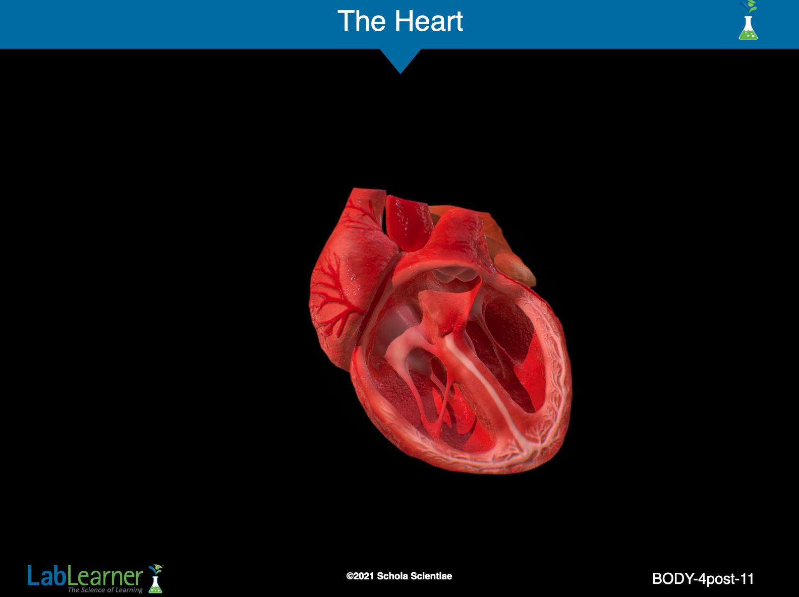
SLIDE BODY-4post-11
Point out to students that blood enters the heart from the body through a major blood vessel called the Superior Vena Cava. After returning to the heart from the lungs, oxygenated blood leaves the heart through the Aorta and circulates through the body before returning to the heart through the Superior Vena Cava once again.
The heart beats close to 100,000 times a day and about 35 million times per year. That is well over 2.4 billion beats in a typical lifetime!
______________________________________________
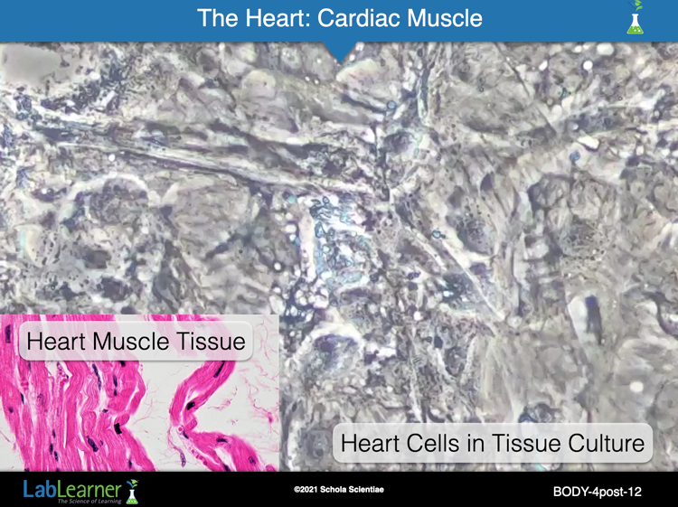
SLIDE BODY-4post-12
In the lower-left of this slide is a section of heart tissue. It is a form of muscle tissue unique to the heart and called cardiac muscle.
The background of the slide shows a video of heart cells grown in a lab as tissue culture. Notice that when heart cells are grown in the lab, they spontaneously contract in a very regular, rhythmic manner. It is awesome to observe!
______________________________________________
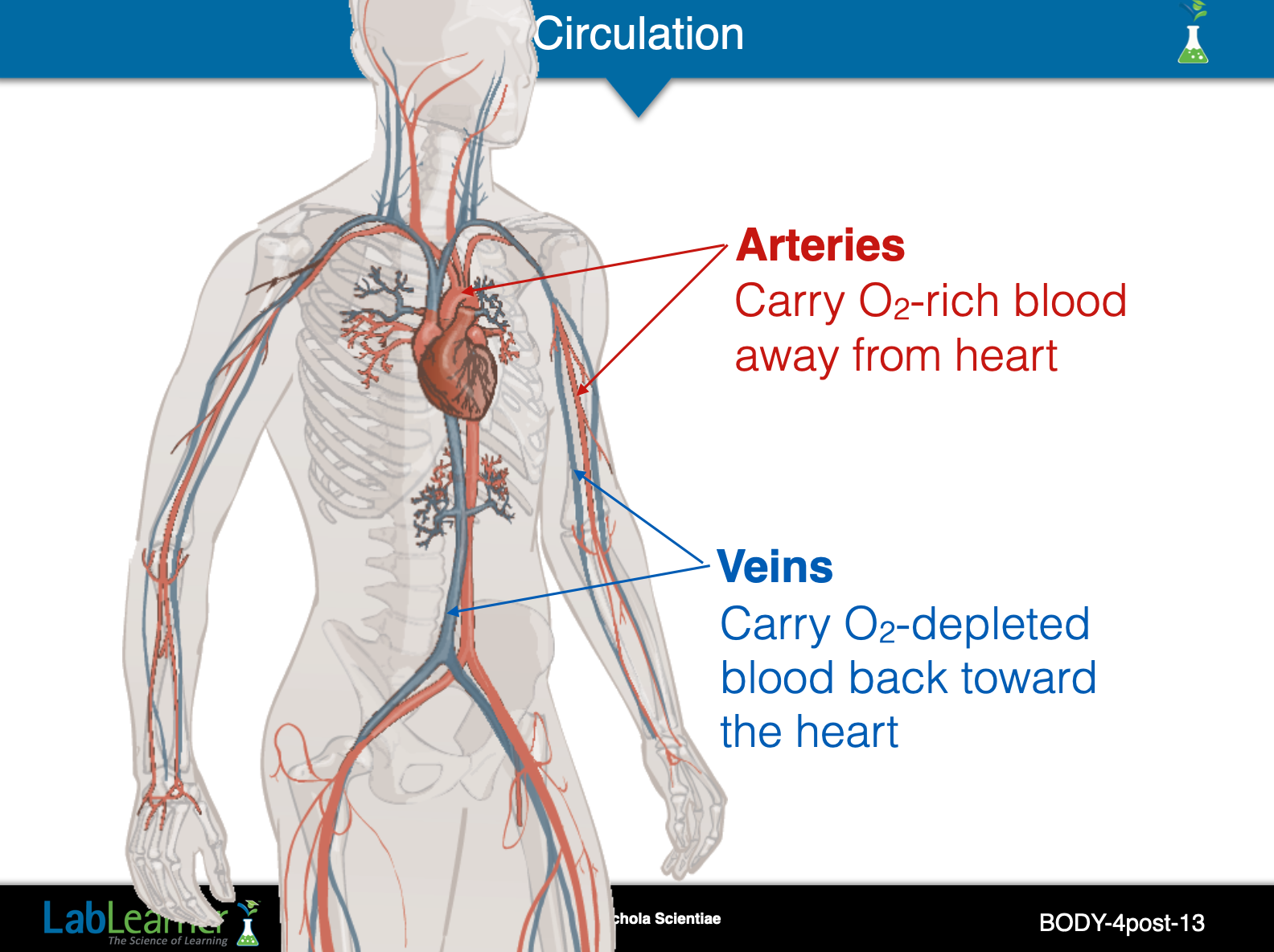
SLIDE BODY-4post-13
Explain to students that the blood moves away from the heart to the rest of the body through blood vessels called arteries. Once the blood reaches the other parts of the body, it moves back toward the heart through blood vessels called veins.
Point out the heart, veins, and arteries to the students (Figure 4.2 in SDRs).
______________________________________________
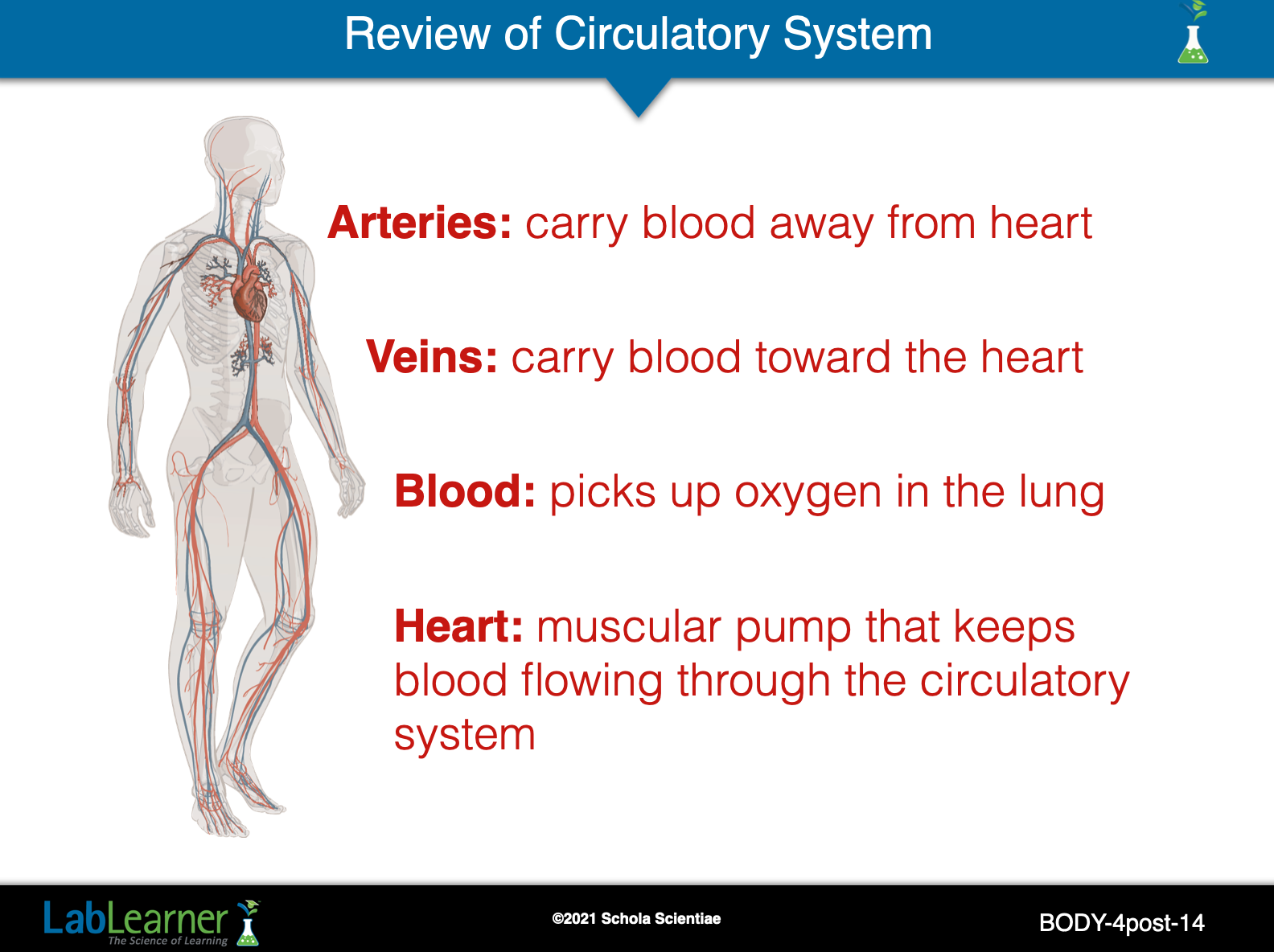
SLIDE BODY-4post-14
This summary slide reviews several key points. It is important to remember that the entire function of the circulatory system is to assure that every cell in the body receives an adequate supply of blood.
______________________________________________