Teacher Portal:
Microscopes and Magnification
Investigation 5
Microscopes and Magnification: Investigation 5
Students began the Microscopes and Magnification CELL by studying the structure of the eye and its functions, such as gathering light, focusing the image, and transmitting this information to the brain, where it is interpreted. In Investigation One the concept of magnification of light by a lens was investigated through experiments using a hand lens.
Investigation Two explored the concept of refraction by different types of lenses, resulting in either an increase or decrease in the apparent size of an object. Student activities focused on the use of lenses to assist the naked eye in increasing or decreasing the resolution of an object’s image. Investigation Three introduced the compound microscope, individual functions of its parts, and how each function contributes to the magnification of microscopic specimens. Students also demonstrated proper techniques for using a microscope, and compared and contrasted its functions with those of a hand lens.
Investigation Four allowed students to further investigate microscopy by investigating the powers of magnification of the three objective lenses and how both resolution and field of view are affected by differing powers of magnification.
In Investigation Five, students will investigate how a microscope functions to refract light by constructing a model of a functioning microscope using hand lenses. Students will, in very general terms, be made aware of how the microscope’s lenses combine to produce an image. The model is a simplified representation of a microscope. It focuses on how the lenses in the eyepiece and objectives of a microscope work. The investigation first explores the effect of refraction by the convex lens in the eyepiece, then focuses on the effect of refraction by the convex lenses in the objectives of the microscope. The final portion of the model combines the refractory effect of the convex lenses in BOTH the objectives and eyepiece.
During prior Investigations, students discovered that the convex lenses found in both the hand lens and in the compound microscope refract the light from an object to produce a magnified image. In addition, students may have also found that the images produced by the hand lens and the microscope were oriented differently. The reason for this lies in the properties of convex lenses as well as in the construction of a microscope.
Microscope construction has changed over the centuries since it’s inception. However, one constant over the course of time has been the use of a lens in a microscope that can refract light in such a way as to produce a magnified image. Many of today’s microscopes use a two lens system with convex lenses in the objectives and another in the eyepiece. This type of microscope is called a compound microscope. However, as with many pieces of scientific equipment, it would be unfair to say that ALL microscopes are constructed exactly the same. What will be described below is one of the most common types of microscope construction and is used here to explain why students may have seen images that appeared differently when using the hand lens and microscope.
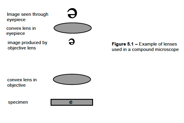 As noted above, many compound microscopes contain a convex lens in each objective and in the eyepiece. However, they produce images that are different because there is a difference in the distance between the object being observed and the lens. The object under observation by the lenses in the objectives is the specimen. The object under observation by the convex lens in the eyepiece is the image produced by the objective. Scientists use a measurement called focal length to describe distances when discussing lenses. For the convex lenses in the objectives, the specimen is between 1 and 2 focal lengths from the convex lens. The image produced by this lens is inverted and magnified. In contrast, the “object” under observation by the convex lens in the eyepiece is LESS THAN one (1) focal length from the convex lens. This produces a magnified and “right side up” image. When combined, the lenses produce an image that is inverted and magnified compared to the specimen.
However, this may or may not be how the specimen actually appears when viewed under the microscope. In addition to lenses, microscopes may also use mirrors or prisms between the objectives and eyepiece to direct the image to the eyepiece. Prisms and mirrors can change the orientation of the image through refraction and reflection. In the end, specimens may appear in the same orientation as on the slide, inverted and magnified, or even inverted, reversed, and magnified. For most scientists working with compound microscopes difference between the specimen’s orientation on the slide and through the eyepiece is not significant. Rather, their attention is focused on the details and observations they can make as a result of the increased resolution the microscope affords.
In Investigation Five, students will build a model of a microscope, focusing on differences in orientation of the specimen and image, and on the contributions that the convex lenses in the objective and eyepiece make to the magnification of the image.
Microscopes and Magnification : Investigation 5 - Mathematics Concepts
PreLab
- Venn Diagram
- Least to greatest
- Greater than/less than/equal to
- Multiplication/division
Lab
- distance in cm
- comparing size, shape, form
- data table
Postlab
- comparing size, shape, form
- angles
- rays
- greater than/less than/equal to
Microscopes and Magnification: Investigation 5 - Procedural Tools

Microscopes and Magnification:
Investigation 5 Quiz

