Teacher Portal:
Microscopes and Magnification
Investigation 5 – Lab
 ASK WHY
Microscopes have made a tremendous contribution to science since their use began in the sixteenth century (the 1500s).
Microscopes are one of the most important scientific instruments developed. In fact, in the medical field, microscopes are largely responsible for making modern medicine “modern”!
BRANCH OUT
Microscopists today work in many different fields including field and laboratory life sciences, chemistry, materials science, and nearly every branch of biomedical research and medicine.
BE PREPARED
Supplies and Equipment:
Group Materials:
- 1 letter “e” slide
- 1 compound microscope
- 2 hand lenses
- 1 meter stick
Individual Materials:
- 1 Student Data Record
Teacher Preparation
- Place required materials at a central location.
- Divide the class into five cooperative groups.
INVESTIGATE
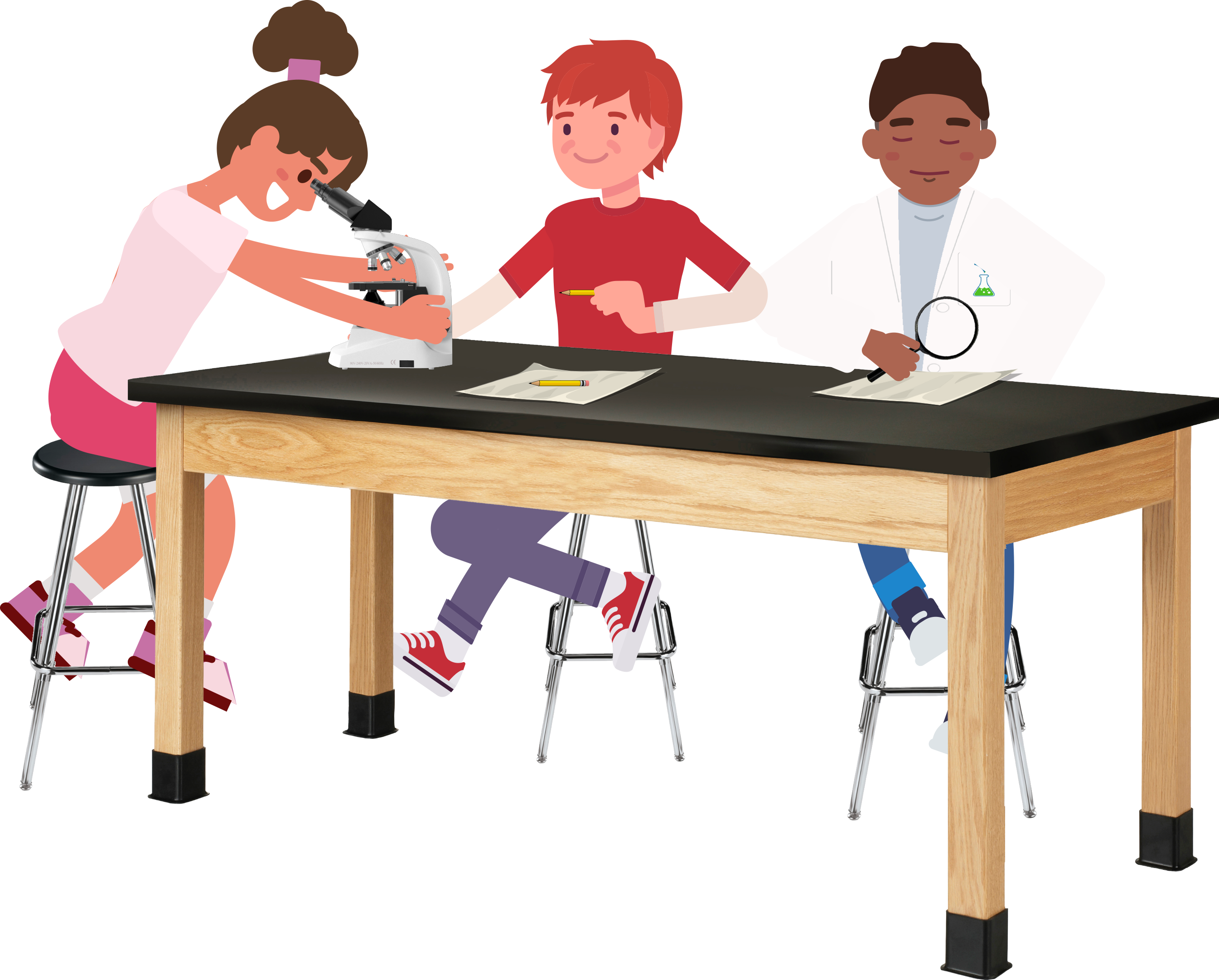
- Direct each student pair to obtain the following required materials: one (1) microscope and one (1) letter “e” slide two (2) hand lenses, and one (1) meter stick.
- Remind students to always hold and carry the microscope with two hands, one on the arm and the other on the base.
1. Inform students that in this Investigation they will compare how their eyes, a hand lens, and a microscope view the image from an object. They will then use a hand lens to model the function of a microscope.
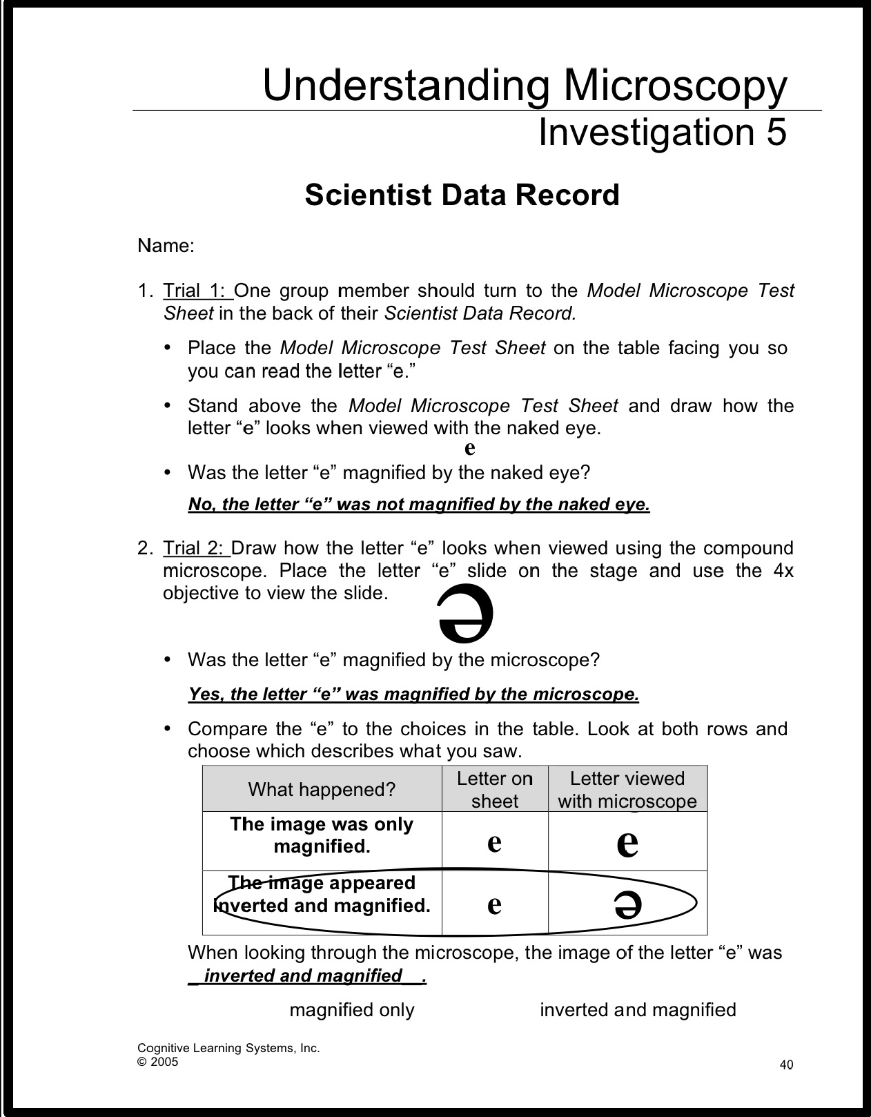
Trial 1:
Inform students that they will first make observations of the image of an object using the naked eye.
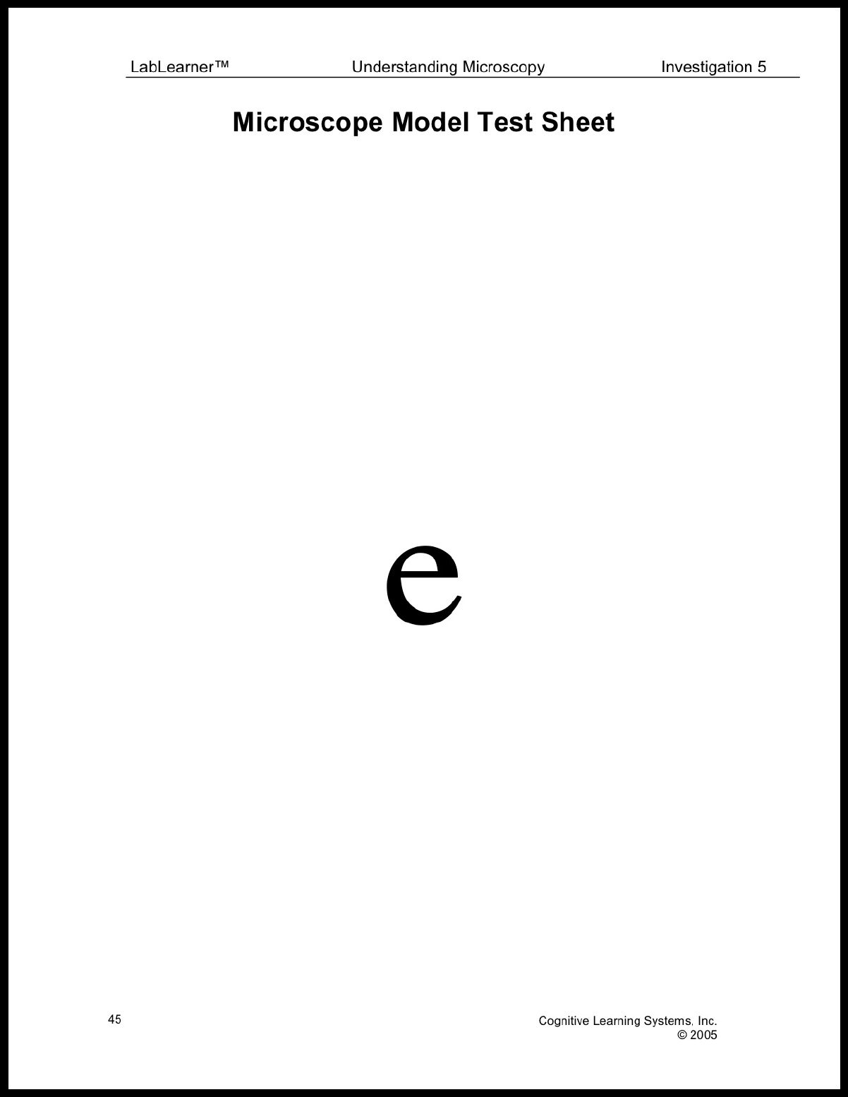
• Instruct a student from each group to turn to the Model Microscope Test Sheet in the back of their Student Data Record.
 • Instruct students to place the Model Microscope Test Sheet on the table facing them so they are able to read the letter “e.”
• Instruct each student in each group to take their turn standing above the Model Microscope Test Sheet and observe how the letter “e” looks when viewed with the naked eye. Students should draw its appearance in Problem 1 in their Student Data Record and answer questions in Problem 1.
Trial 2:
Inform students that they will next make observations of the image of an object with the microscope.
• Instruct students to place the slide under the clips on the stage of the microscope and use the low-power objective to view the slide. Students should draw the appearance of the letter “e” in problem 2 in their Student Data Record.
• Students should answer the questions in Problem 3 in their Student Data Record.
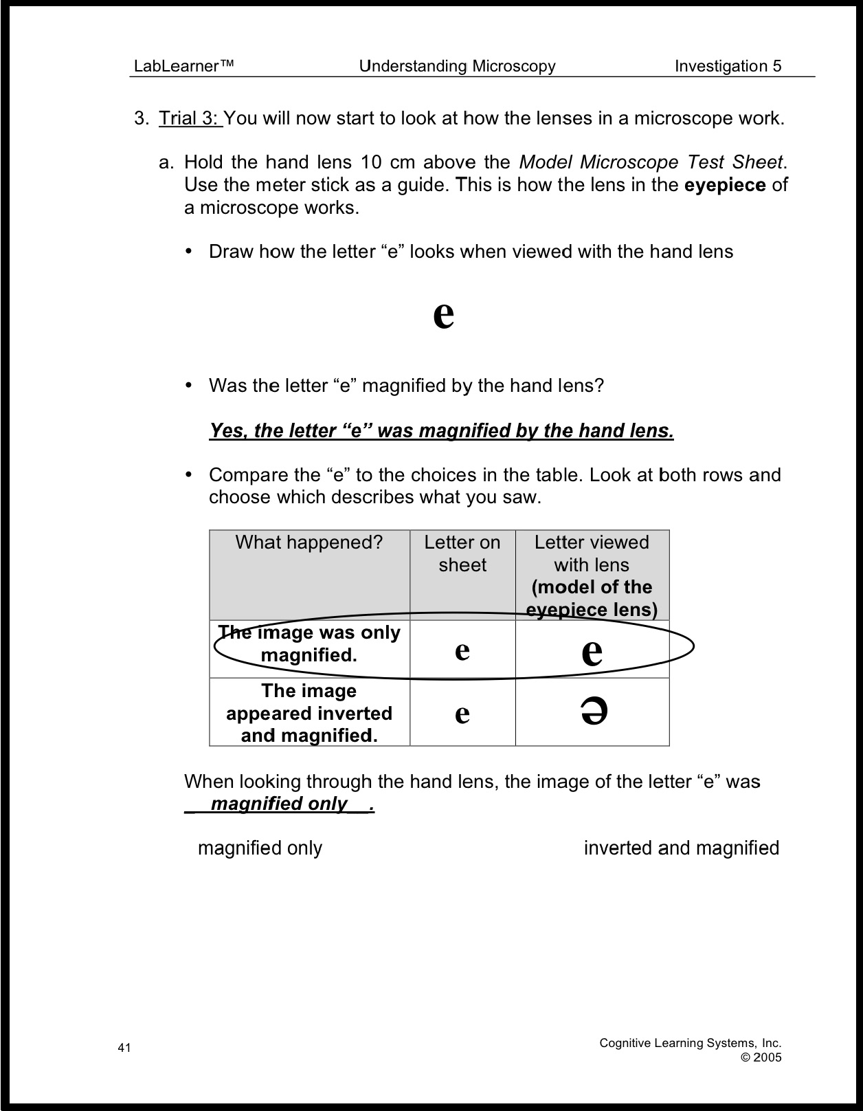
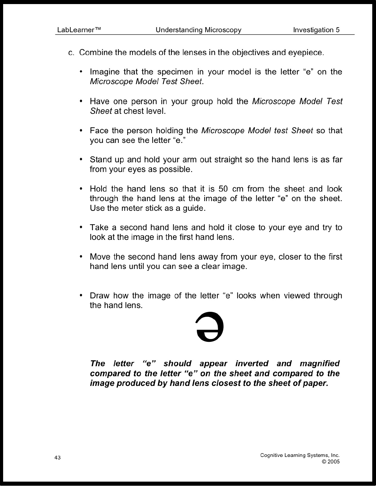
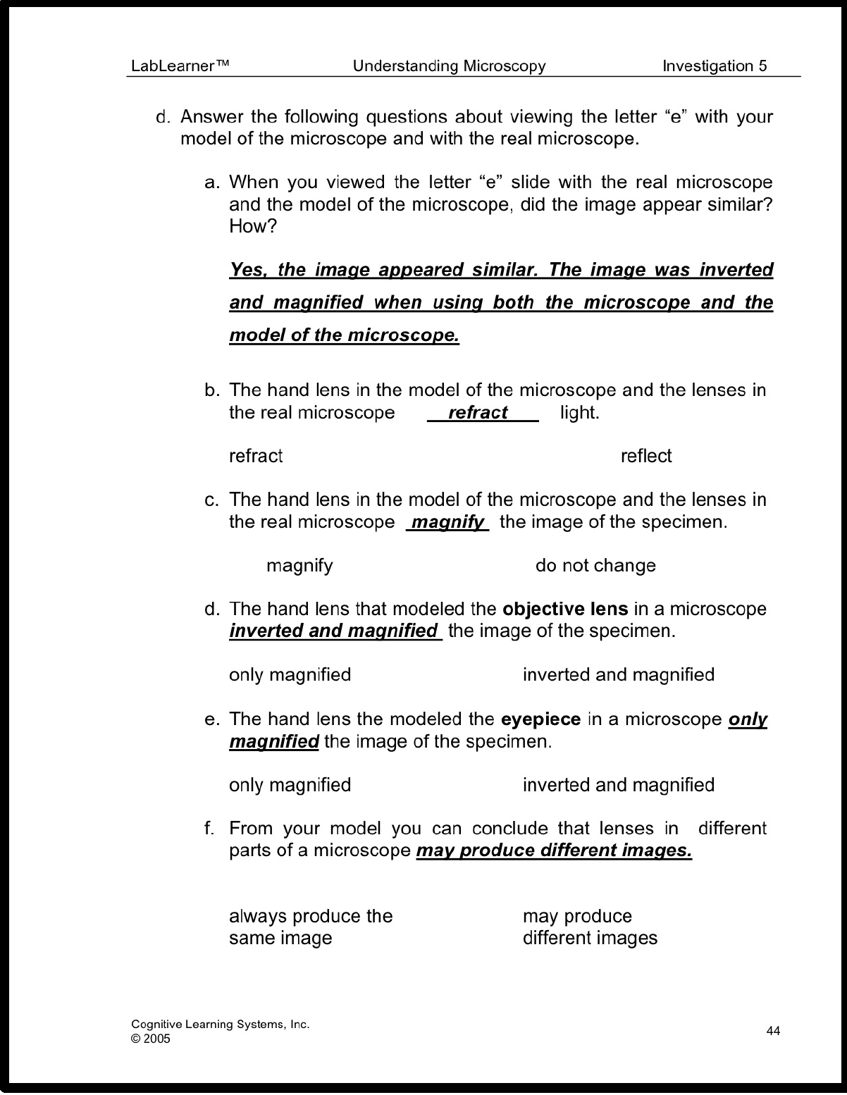
Trial 3
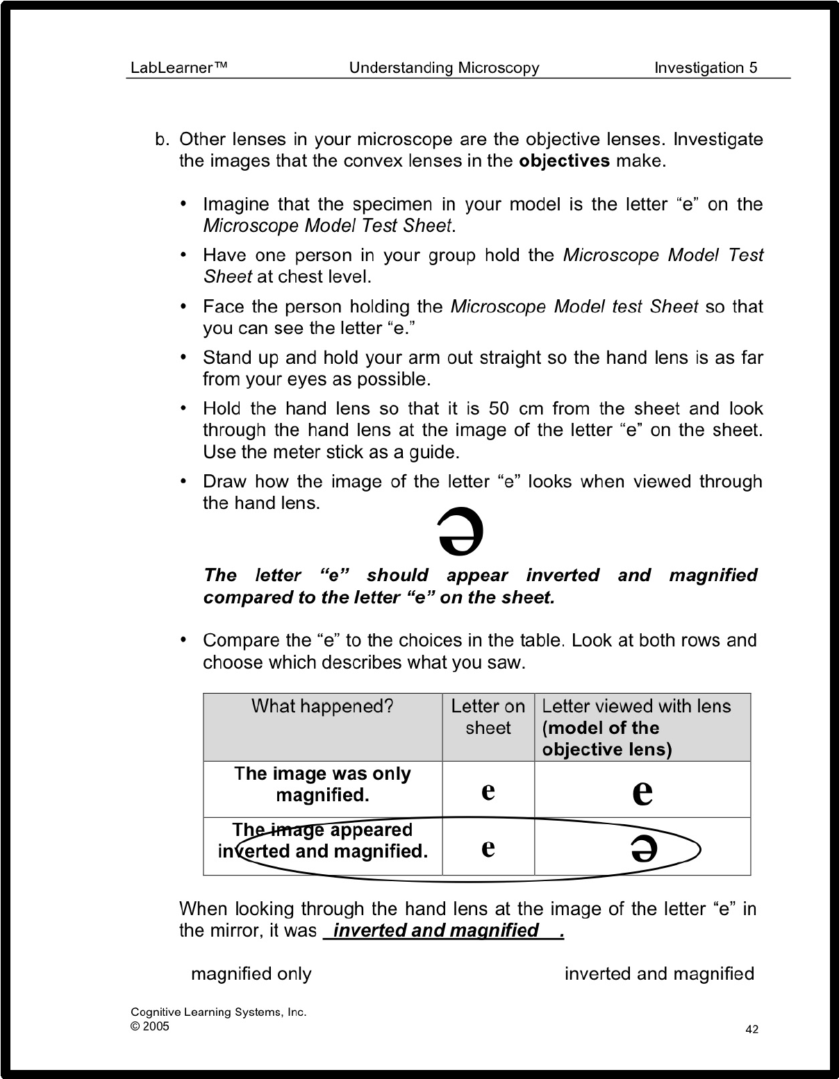
Inform students that they will investigate how the lenses in a microscope work. During this trial, students will model the image formed by the convex lenses in the objectives of a compound microscope and the convex lens in the eyepiece of the microscope.
- To begin the Trial, students will first model the image produced by the convex lens in the eyepiece. Once completed, students will then investigate the image produced by the convex lens in an objective of the microscope.
- Finally, students will model the image formed by the combination of the convex lenses in the objectives and eyepiece. Students should find that the convex lenses in both the eyepiece and objective lenses produce a magnified image. However, the image produced by the convex lenses in the objectives also inverts the image, whereas the convex lens in the eyepiece does not. This finding should help students understand that a convex lens has the ability to produce different types of images and that understanding these differences is helpful in understanding the resolving power and appearance of images produced by the microscope.
KEYS

CLEAN UP
Let students know your expectations for clean-up. Ask them to clean up.