Teacher Portal:
Microscopic Explorations
Investigation 3
Microscopic Explorations: Investigation 3
Students’ explorations during Investigations One and Two provided opportunities for them to better understand the effects of the refraction of light through lenses and how these effects can be used to provide different magnifications, resolutions, and fields of view when studying specimens.
In Investigations Three, Four, and Five, students’ focus shifts from the investigation of refraction, lenses, and the operation of the microscope to the observation and analysis of cells and tissues. Students begin their analysis in Investigation Three with an introduction to animal cells.
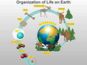 First, it is important to consider exactly where cells fit into the scheme of the overall organization of life on Earth. The illustration shown here depicts the organization of life from the atomic level, near the bottom, to the entire biosphere, which is the sum total of all life on Earth.
Along this path of organization, atoms bind to each other to form molecules. Molecules are then organized into the membranes and organelles that are found in cells and will be discussed shortly. All the trillions of molecules, membranes, and organelles come together to form the most fascinating and basic unit of life, the cell. Many, many millions of years  ago, this is where the organization of life on Earth stopped. That is, there was a time when only single-cell life existed on our planet. However, the organization of life continued to form more complicated structures by the interaction of single cells into tissues and individual multicellular organisms, like humans and trees. The further organization of individuals into populations and ecosystems will be the topic of many other LabLearner CELL units.
Not all single-cells evolved into multicellular organisms. Even today, there are numerous, countless species of single-celled organisms including bacteria that thrive on the planet. In addition, there is a multitude of multi- cellular organisms such as plants and animals that exist along with them. In general, a cell is described as the smallest part of a living organism that can support life and in multicellular organisms is considered the organism’s basic unit of structure and function. Although most cells are between one and one hundred micrometers in diameter (1000 micrometers = 1 millimeter) cells also contain smaller parts referred to as organelles.
One of the most interesting concepts about cells is that although cells in different organs and organisms can be very different in their structure and function, many of the organelles within cells are the same.
Therefore, differences in cell structure can be explained as the result of variations in cellular organelles, and many differences in cell function can be attributed to the differences in cell structure. Ultimately the differences in both structure and function are the result of the instructions located within the DNA of each cell. For example, all cells have a cell membrane (or plasma membrane) or outer shell that serves as the boundary of the cell. The cell membrane helps to regulate substances that can enter or exit the cell. However, the membrane of different cells may allow different substances to enter and exit the  cell. The cell membrane of a heart cell, for example, operates slightly differently than the cell membrane of a kidney cell.
In this CELL students will focus their investigation on cells from plants and animals. Because the basic parts of cells are similar, when first introduced to cells students can benefit from studying a basic model of a cell. As students progress with their science education, they can build upon this model to understand both the multitude of differences and similarities in cell structure and function that exist in cells from different tissues, organs, and organisms. Therefore, in this CELL students will study a basic model of an animal, plant, and bacteria cell.
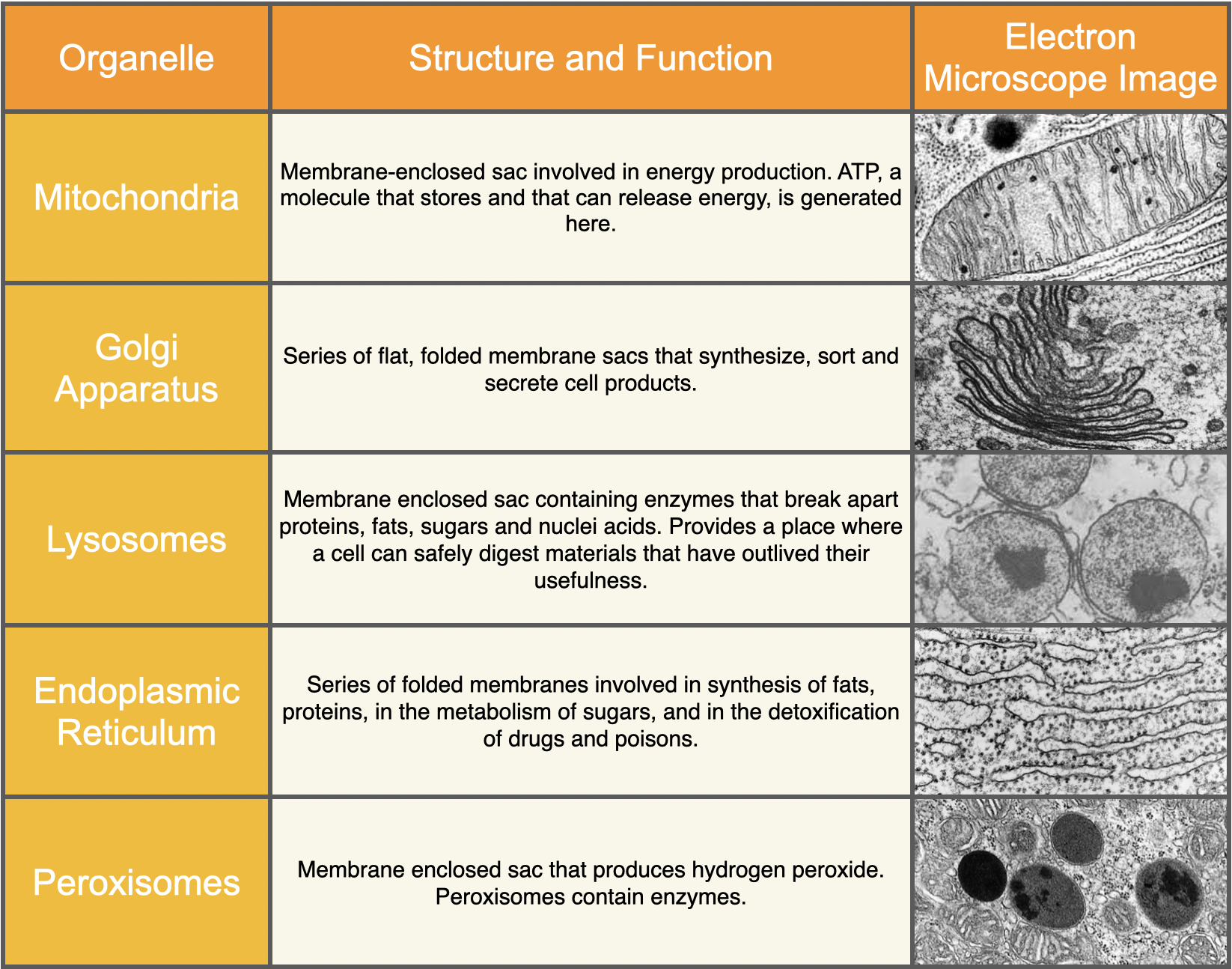
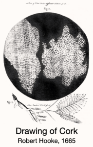 Cells were discovered in 1665 by Robert Hooke. However, much of what we know about the parts of cells did not come until the 1950s with the advent of the electron microscope. From observations with the electron microscope, scientists found there are several basic components that most cells have in common: cell membrane, cytoplasm, chromosomes (DNA), and ribosomes. As noted above, the cell membrane functions as a selective barrier for the cells, regulating substances that both enter and exit the cell. The cytoplasm is a semi-fluid or gel-like substance located within the boundaries of the cell membrane. Most other parts of the cell are located within the cytoplasm. Chromosomes, the structure consisting of protein and DNA, serve to regulate cell function and structure through the instructions located within the DNA. Ribosomes have a special purpose as they help “translate” the instructions in DNA into products the cell needs to function. In addition to these basic organelles, other organelles that are commonly found in animal cells, the cells that students will observe in Investigation Three, include mitochondria, Golgi apparatus, lysosomes, endoplasmic reticulum, and peroxisome. The table below provides a summary of some of the primary functions of each of these organelles.
In contrast to the cell membrane, cytosol, (or cytoplasm), and nucleus, these organelles are not easily resolved or visible with the compound microscope.
Although many cells share the same parts, cells can be classified according to their similarities and differences. Two such classes of cells include eukaryotic cells and prokaryotic cells. In eukaryotic cells, the chromosome (DNA) is contained within a membrane-enclosed sac called the nucleus. Cells found in plants and animals are considered eukaryotic cells. In a prokaryotic cell, chromosomes (DNA) 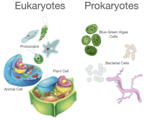 are located within one region of the cell but are not separated from the rest of the cell by a membrane. Although prokaryotic cells also lack some of the other organelles found in eukaryotic cells, this difference in nuclear structure is one of the most frequently used criteria for differentiation between the two. A second is size. Prokaryotic cells are generally much smaller than eukaryotic cells. For example, most bacteria, which are prokaryotic, are 1- 10 micrometers in diameter. Eukaryotic cells, generally range from 10-100 micrometers in diameter.
As a way of learning about cells and their organelles, students will prepare a specimen of their own cheek cells. In doing so, they will use a new Procedural Tool: Wet Mount Slide Preparation. Observation of their cheek cells with the compound microscope will give students a hands-on opportunity to view the cell membrane, cytoplasm, and nucleus of a cell.
Microscopic Explorations: Investigation 3 - Mathematics Concepts
Prelab
• greater than/less than/equal to
• (in)direct relationships
• multiplication/division
• problem solving
• predicting/verifying size, shape, form
• parts/whole
Lab
• multiplication
Postlab
• data table
• sequential order
• multiplication/division
• length/height/width in cm
• counting whole numbers
• greater than/less than/equal to
• comparing (non)measurable characteristics
• diameter in mm/cm
• metric conversion
Microscopic Explorations: Investigation 3 - Procedural Tools

Microscopic Explorations:
Investigation 3 Quiz

