Teacher Portal:
Microscopic Explorations
Investigation 3 – PostLab
ASK WHY
Microscopes are one of the most important scientific instruments developed. In fact, in the medical field, microscopes are largely responsible for making modern medicine “modern”!
BRANCH OUT
You might wonder which came first, the microscope or the telescope. Actually, they appear to have appeared at about the same time although it is thought that the microscope may have come first. In either case, the first forms of both instruments were developed in the 16th century (the n1500s). It isn’t that much of a stretch to imagine that, once the microscope was invented, that someone would consider making similar instruments that could make distant objects appear closer.
PRINT IT
Use your browser to download a printable PDF as help during the slide presentation and to make additional notes. In your browser, go to File > Print and then choose to save as PDF.
NAVIGATE IT
Once the slide presentation is launched
- use your left and right arrows to advance or go back in the slide presentation, and
- hover your mouse over the left edge of the presentation to get a view of the thumbnails for all the slides so that you can quickly move anywhere in the presentation.
- Click HERE to launch the slide presentation for the CELL.

Note to Teacher: Out of an abundance of caution, do not have your students perform step #7 in your PostLab 3-ring Teacher Manual. This step asks students to look in each other’s mouths. We should hopefully return to this interesting activity next year.
SHARE IT
SLIDE MICRO3-post-1
We will begin the PreLab with a review of the experiments students performed in the lab.
______________________________________________
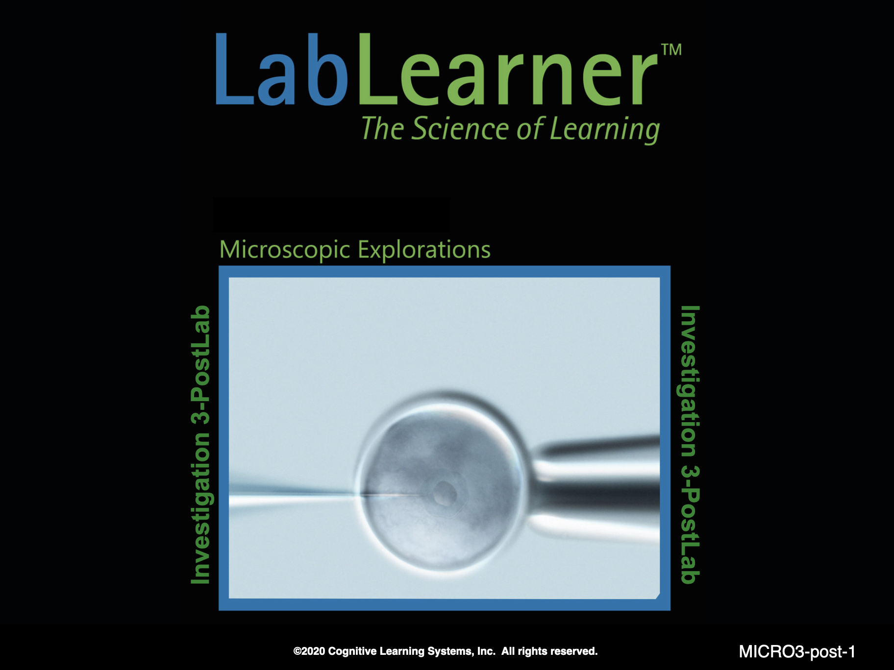
SLIDE MICRO3-post-2
Begin this part of the investigation by reviewing the experiments students performed in the lab. Ask the following questions to promote discussion of the experiments.
Ask students: What was the theme or focus of the investigation? The theme or focus of the investigation was to examine how cells from living tissues appeared under the microscope at varying powers of magnification. Students investigated the appearance of animal cells by preparing a specimen of their cheek cells and observing the cells using the compound microscope.
Remind students that in Trial 1, they prepared a wet mount slide. Ask: What questions did you answer when performing Trial 1? How did you answer this question? Students investigated the questions: What is a wet mount slide? What is the procedure for making a wet mount of cheek cells? To answer this question, students obtained a sample of cells from their cheeks and followed the steps described in their Scientist’s Data Record for making a wet mount slide.
Remind students that in Trial 2, students used the microscope to observe the wet mount slide that they made. Ask: What question was Trial 2 designed to answer? How did you answer this question? Trial 2 was designed to answer the question: What information about the structure and function of cheek cells can be learned from microscopic analysis? To answer this question, they viewed a sample of their cheek cells using the three objectives of the compound microscope.
______________________________________________
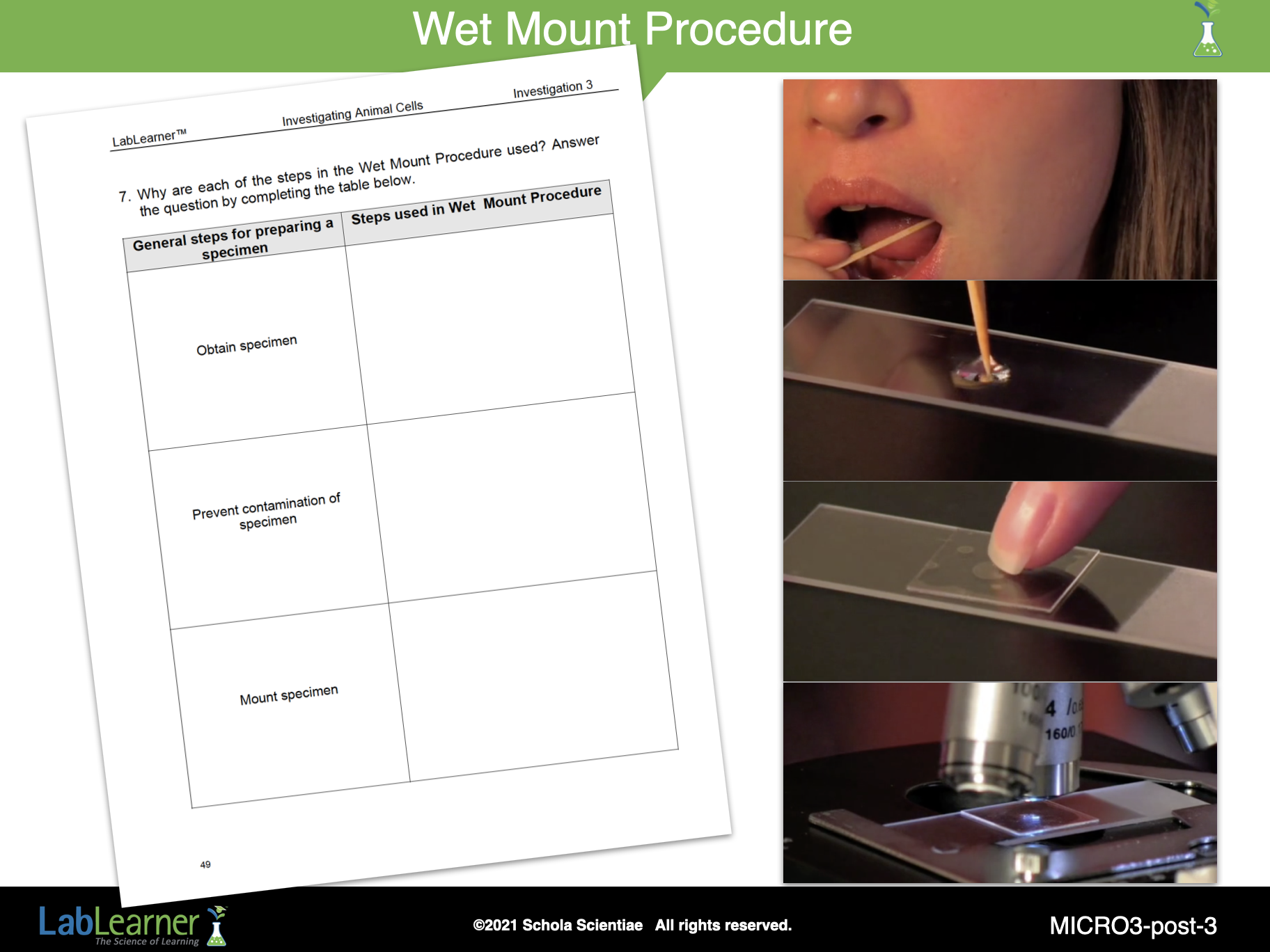
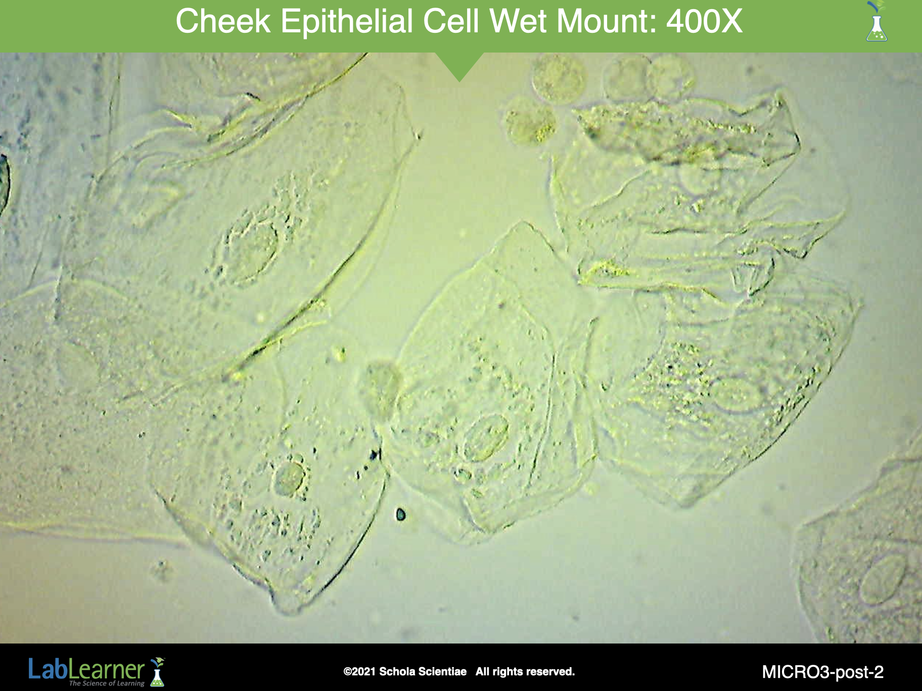
SLIDE MICRO3-post-3
Help students compare the steps they performed in Investigation Two with those they used to prepare the wet mount of their cheek cells.
a. Review the steps students performed in Investigation Two and the importance of each step. Draw students’ attention to the general descriptions that can be used to categorize each of the steps.
b. Divide students into groups of five and assign them the task of matching the steps they used in preparing a wet mount to the general descriptions for specimen preparation. Students should record their answers in Problem 7 of their Scientist Data Record.
c. Once students have completed their discussions within their groups, ask one person from each group to share their answers. Students can record the answers in the last column of the Table in Problem 7.
______________________________________________
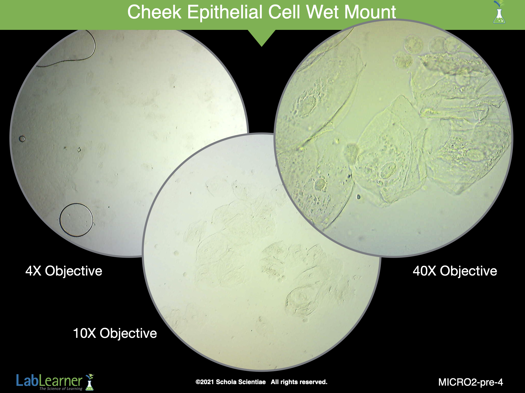
SLIDE MICRO3-post-4
Continue the analysis of the investigation by discussing students’ observations of Trial 2: microscopic analysis of their cheek cells.
Ask students: Which objectives did you use to view the cheek cells? Students viewed the cheek cells with the low (4X), medium (10X) and high (40X) power objectives.
Ask students: What was the power of magnification when the specimens were viewed with the three different objectives? How did you calculate the power of magnification? Students should answer that the power of magnification was calculated by multiplying the magnification power of the objective by the magnification power of the eyepiece. Both the power of magnification of the objective and the eyepiece were included in the equation because there is a lens in both the eyepiece and the objective of the microscope. Each lens has a refractive power. Therefore the total magnification is the product of the magnification of both lenses.
Total power of magnification of the low (4X) objective
Objective power X eyepiece power
4 X 10 = 40X
Total power of magnification of the low (10X) objective
Objective power X eyepiece power
10 X 10 = 100X
Total power of magnification of the low (40X) objective
Objective power X eyepiece power
40 X 10 = 400X
Ask students: Describe some of your observations of the cheek cells when viewed through the medium and high power objectives. Which structures and organelles of the cells did you observe? Student answers will vary. However, students’ answers should include a description of the cheek cells under each viewing condition. Students should indicate that the cell membrane, cytoplasm, and nucleus could be seen through both the medium and high power objectives.
Ask students: What do your observations tell you about cheek cells? Are they eukaryotic or prokaryotic cells? How do you know? Students should indicate that cheek cells are eukaryotic cells because they possess a clearly defined nucleus. The nucleus of each cheek cell was visible when viewed with the microscope.
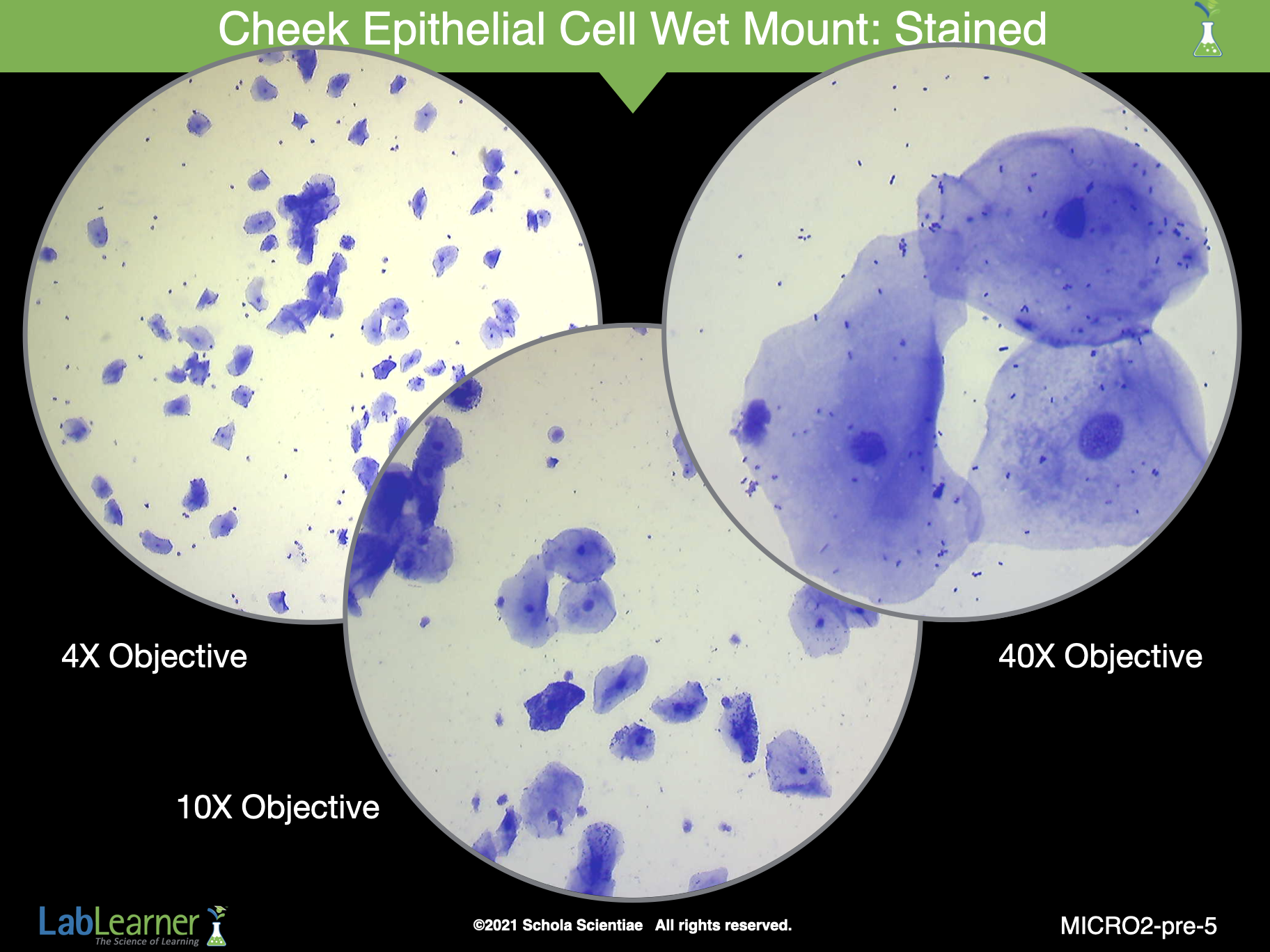
SLIDE MICRO3-post-5
This slide shows check cells that were stained with color.
Ask students: Explain any differences in the appearance of the cells when the two objectives were used. Use the sketches you drew in your Scientist Data Record to help you. Student answers will vary. However, students should indicate that as the magnification power of the objectives increased from 10X to 40X, the cells were magnified and it was easier to see the detailed structure of the cells. For example, the nucleus of each cell was visible when viewed with the medium power objective. However, it appeared to be a small dot. When viewed with the high power objective, it was easier to see that the nucleus was a small oval or round sac that was colorless.
Ask students: You described the differences in the specimens when viewed with the two objectives. Did you observe any similarities in the images of the cheek cells when they were viewed with the three objectives? Can you describe the similarities? Student answers may vary. However, student answers should discuss the following similarities: the color and proportions of the cheek cells were similar when viewed with the two objectives. In other words, the different lenses did not produce a change in color or a change in the proportions of the images.
______________________________________________
POSTLAB EXERCISE
Exercise 1
Divide students into five groups. Distribute two pieces of plastic wrap and a plastic cup with water to each group. Direct students to find Problem 8 on their Scientist Data Record and read the instructions for a new activity.
Once students have completed the activity, discuss their conclusions.
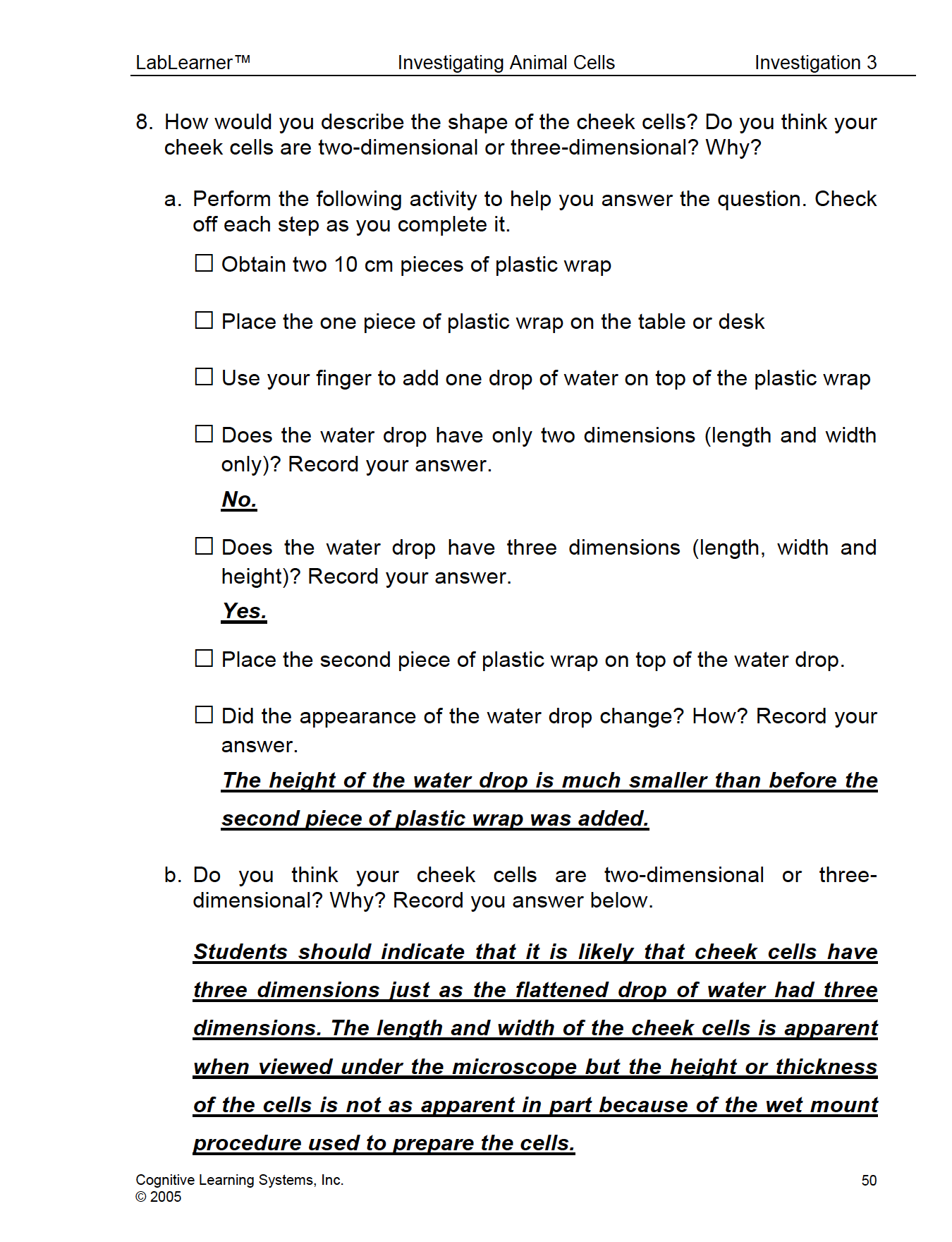
How many dimensions did the water drop have when it was placed on the piece of plastic wrap? The water drop had three dimensions. It had a width and length, which were likely the same and it had a certain height.
What happened when the second piece of plastic wrap was added on top of the water drop? Did the water drop still have three dimensions? How did its appearance change? The water drop was rounded before the second piece of plastic wrap was added. When the plastic wrap was placed on top, the water drop was flattened. It still had three dimensions: width, length, and height. However, its height was much less than prior to its covering with the plastic wrap. When the water drop was flattened, it appeared to have only two dimensions: width and length.
Was the activity with the plastic wrap similar to a procedure you performed in the lab? Which one? The activity with the plastic wrap was similar to the wet mount procedure used to prepare the cheek cells for microscopic analysis.
Do you think cheek cells have two dimensions or three dimensions? Why? Students should indicate that it is likely that cheek cells have three dimensions just as the flattened drop of water had three dimensions. The length and width of the cheek cells are apparent when viewed under the microscope but the height or thickness of the cells is not as apparent in part because of the wet mount procedure used to prepare the cells.
Exercise 2
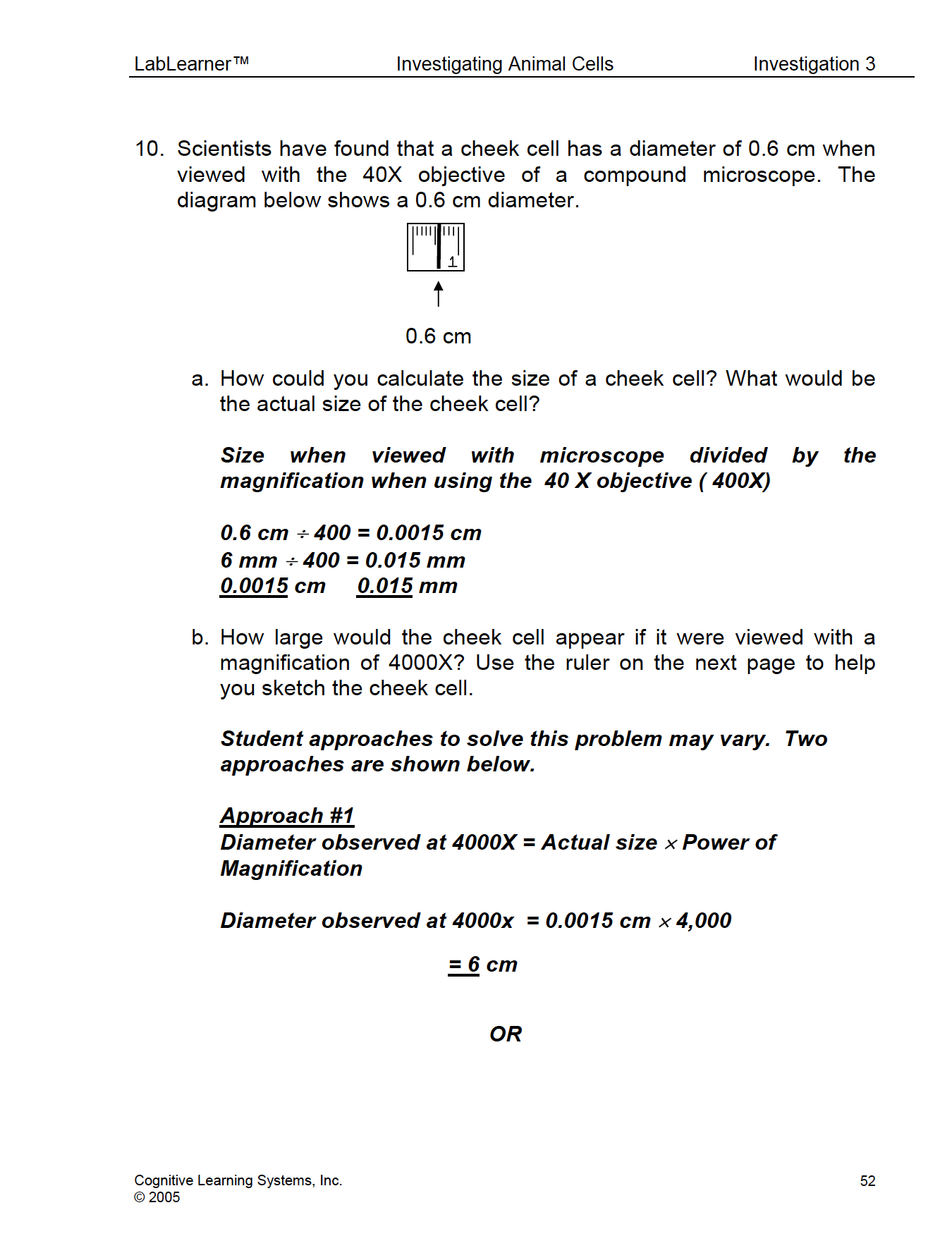
Direct students to find Problem 10 in their Scientist Data Record. Explain that when viewed with the 40X objective, the width of the image of the cheek cell is approximately 0.6 cm or 6 millimeters.
Encourage students to work with a partner to complete Problem 10a in their Scientist Data Record and determine the actual size of a cheek cell.
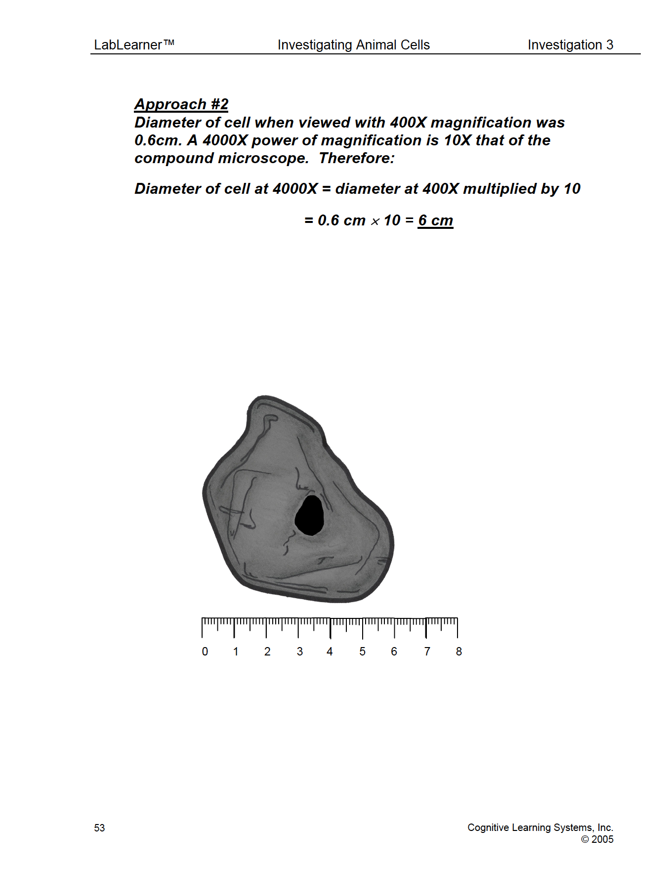
Challenge students to apply what they have learned about the power of magnification and drawing objects to scale by answering Question 10b in their Scientist Data Record. In this question, students are asked how large the cheek cell would appear if it were viewed with a magnification of 4000x.
End Investigation Three by reminding students that their exploration of cells has just begun. In Investigation Four students will prepare and study examples of cells from plants. In preparation for the next investigation ask students:
- Do you think plant cells are different than animal cells? Why? How could you find out?
- Do you think plant cells are similar to animal cells? Why? How could you find out?
KEYS: POSTLAB EXERCISE