Teacher Portal:
Microscopic Exploration
Investigation 3 – PreLab
ASK WHY
Microscopes are one of the most important scientific instruments developed. In fact, in the medical field, microscopes are largely responsible for making modern medicine “modern”!
BRANCH OUT
Cell Biologists study how cells work. Cell Biologists are involved in important research in many different areas of biological sciences. They are employed at universities, hospitals, clinics, and industry.
PRINT IT
Use your browser to download a printable PDF as help during the slide presentation and to make additional notes. In your browser, go to File > Print and then choose to save as PDF.
MINDSET
This Investigation is designed to:
- introduce students to the concept of a cell as the smallest unit in an organism capable of sustaining life.
- introduce students to examples of eukaryotic and prokaryotic cells.
- provide students an opportunity to view an example of a eukaryotic animal cell (cheek cell) with the compound microscope.
- provide students an opportunity to prepare a specimen for microscopic analysis and to learn a new Procedural Tool: Wet Mount Slide Preparation.
- help students understand the relationship between the structure and function of cells and their parts.
- review the concept of scale and the importance of accurately representing microscopic observations.
- promote the use of differences in the field of view, magnification and resolution of the low, medium and high power objectives of the compound microscope when observing specimens.
SCIENTIST’S GLOSSARY
- Cell: the smallest structure of living organisms that can perform functions necessary for life. Most cells are microscopic.
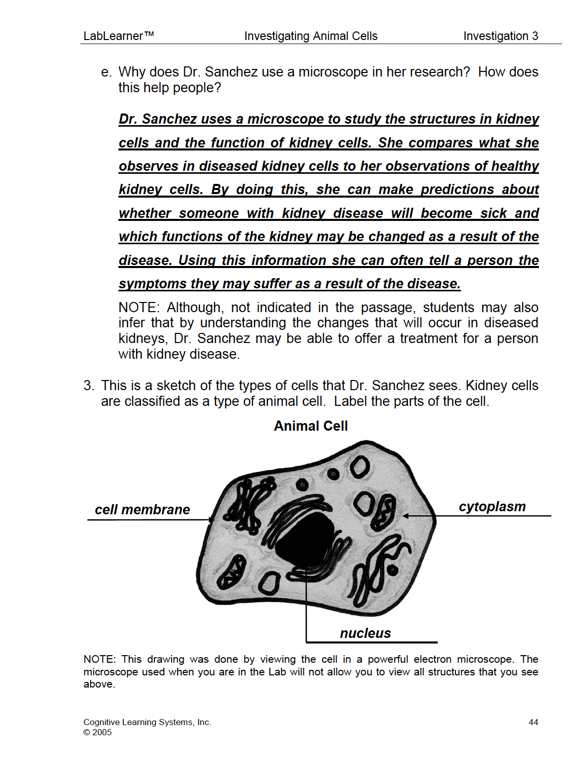
- Cell membrane (Plasma membrane): the outer layer of a cell. The cell membrane helps control what can enter and exit a cell.
- Cytoplasm: the gel-like area of a cell between the nucleus and cell membrane where many of the functions of the cell occur.
- Eukaryotic cell: a cell that contains a defined nucleus.
- Nucleus: the small sac inside a cell that contains or contained DNA; one type of organelle in a cell.
- Organelle: a small structure inside a cell that performs a specific function.
- Prokaryotic cell: a cell that does not contain a clearly defined nucleus.
BE PREPARED
Watch the Investigation 3 Teacher Video and Student Video below to prepare for the PreLab.
SET FOR SUCCESS
- Tell students that they are about to begin their study of animal cells.
- Ask students to share the kinds of things they expect they might learn in these Investigations.
- Tell students they will use the compound microscope to examine cells, including their own.
Begin the PreLab Concept Slides to start students on their learning journey. Then watch the Pre-Lab Student Video as a class.
NAVIGATE IT
Once the slide presentation is launched
- use your left and right arrows to advance or go back in the slide presentation, and
- hover your mouse over the left edge of the presentation to get a view of the thumbnails for all the slides so that you can quickly move anywhere in the presentation.
- Click HERE to launch the slide presentation for the CELL.
SHARE IT
SLIDE MICRO3-pre-1
In Investigation Three, students will learn about animal cells. In the process, they will learn to make a wet mount microscope slide of their own cheek cells and observe the nuclei of these cells in a compound microscope.
In addition, students will learn about sub-cellular structures called organelles and the differences between eukaryotic and prokaryotic organisms.
______________________________________________
SLIDE MICRO3-pre-2
This slide provides a review of Investigation One and Two:
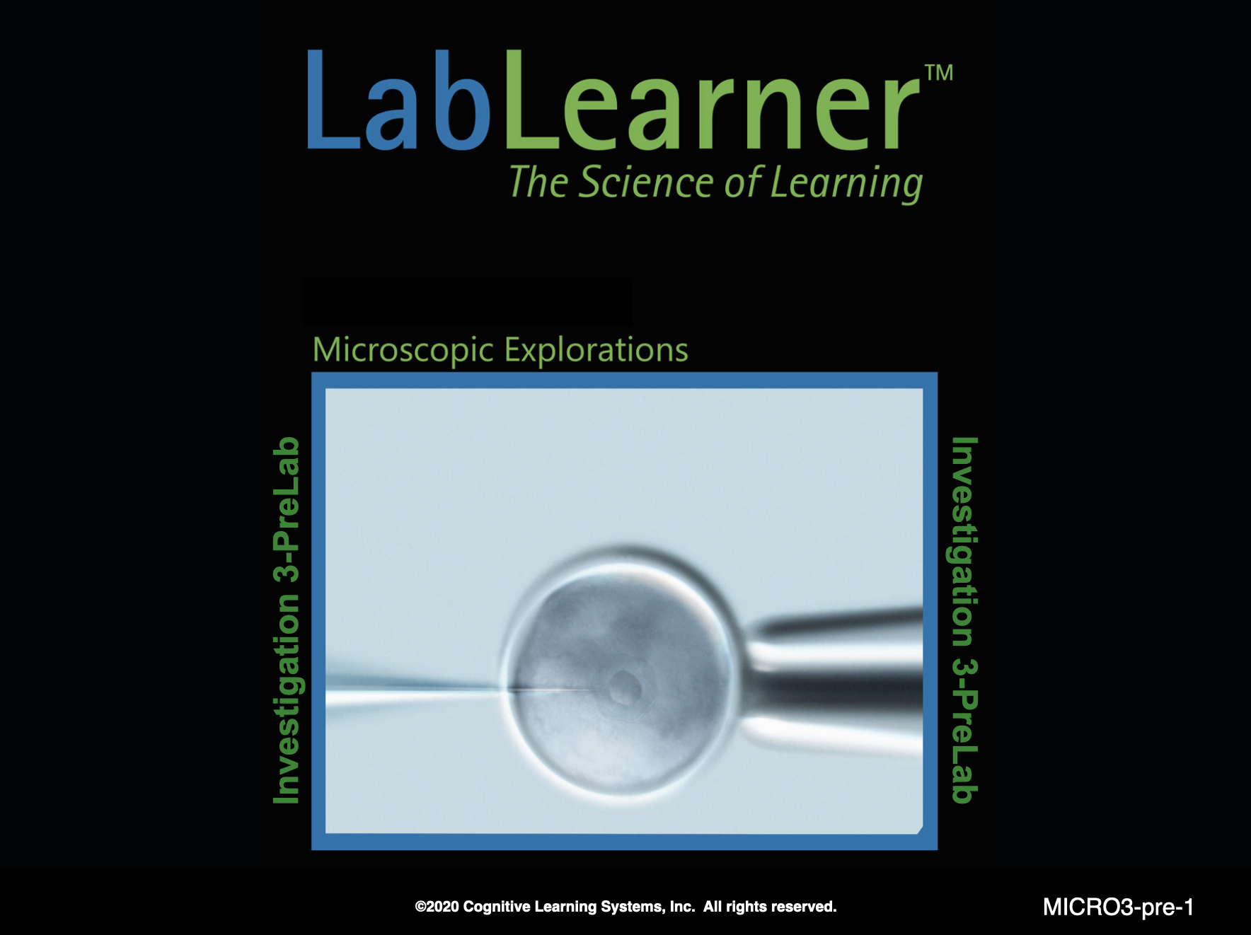
What happens to light when light is refracted? How does refraction change the image of an object? Light rays are bent when light is refracted. An image of an object results from the refraction of light. The image often takes on a different appearance than the object. For example, a concave lens refracts light. The image seen with the concave lens is reduced in size compared to the object.
Can you give some examples of different types of lenses? How are they different from one another? Examples of lenses include convex and concave lenses. Convex lenses are thicker in the middle than at either end. Concave lenses are thinner in the middle than at either end.
How do lenses affect the resolution of the images of an object? Lenses can produce images that have both increased and decreased resolution as compared to the objects viewed. Convex lenses often produce images that have increased resolution as compared to the object viewed. Concave lenses produce images that have decreased resolution as compared to the object viewed.
To help answer this question, “Which lens in this slide do you think would produce the most magnified image? The least magnified image?”, discuss the following question:
Think about what you know about the refraction of light. Would a piece of glass refract light? Why? Students should indicate that a piece of glass would refract light because refraction occurs when light rays travel from one transparent medium to another. Light passing from air to glass is an example of light traveling from one transparent medium to another.
Think about what you see when you look out a window. Do the objects you see look different than when you view them without looking through a window? Students should indicate that in most circumstances, the objects appear similar when viewed through a pane of glass in a window and when viewed without a pane of glass.
Think about what you observed when you look at objects in Investigation One through a convex and concave lens. Did the images you see appear different than the objects? Students should indicate that the images they viewed through the convex and concave lenses appeared different from the object as seen with the unassisted eye.
What is the difference in shape between a pane of glass and the convex and concave lenses you used in Investigation One? Student answers may vary. Help students understand that one of the differences in the shape of lenses versus a pane of glass. A pane of glass is flat whereas the convex and concave lenses are curved pieces of glass.
The ability of a substance to refract light is called the refractive power. Which would have greater refractive power, a pane of glass or a convex lens? How does refractive power affect the types of images seen by the pane of glass and the convex lens? Students should infer that because the images of objects appear different when viewed through convex and concave lenses, but often do not appear different when viewed through a flat pane of glass, that more refraction occurs as light passes through the convex and concave lenses than the flat pane of glass. Therefore, the lenses have more refractive power than the flat pane of glass. The refractive power of the concave lens produces images that are reduced in size as compared to the object. The convex lens can produce images that are magnified as compared to the original object. The lower refractive power of a flat pane of glass results in images that appear very similar to the original object.
Look at the three examples of lenses on this slide. Which of the lenses would have the greatest refractive power? Students’ answers will vary. Students should be able to infer from their experiments in Investigation One and through the series of answers to the preceding questions that the lens with the greatest curvature would have the greatest refractive power.
Which of the lenses would produce the most magnified image? The least magnified image? Students should indicate that all of the images above illustrate convex lenses. Based on their experience in using a convex lens in Investigation One, students should indicate that convex lenses produce magnified images. Therefore, students should conclude based on their experiments from Investigation One and their answers to the preceding questions that the convex lens with the greatest curvature is most likely to produce the most magnified image. The convex lens with the least curvature of the three is most likely to produce the least magnified image.
What do you know about the curvature of a lens and its refractive power? The greater the curvature of a lens, the greater its refractive power.
______________________________________________
SLIDE MICRO3-pre-3
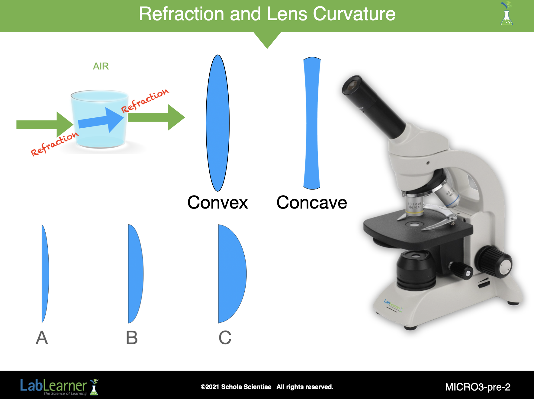
What is the relationship between the resolution of an image, the field of view of an image, and the magnification of the lens used to view an object? As the power of magnification of a lens increases, the resolution of images increases but the field of view decreases. In other words, as the power of magnification increases, more details of the object can be seen, but less of the object is visible under the lens.
Explain one way you could calculate the magnification power when using an objective of the compound microscope. The power of magnification can be calculated by multiplying the magnification power of the objective by the magnification power of the eyepiece. Both the power of magnification of the objective and the eyepiece are included in the equation because there is a lens in both the eyepiece and the objective of the microscope. Each lens has refractive power. Therefore the total magnification is the product of the magnification of both lenses.
Total power of magnification of the low (4X) objective
Objective power X eyepiece power
4 X 10 = 40X
What is the benefit of viewing a specimen with a large field of view versus a high resolution? Student answers may vary. Student answers should reflect the understanding that a large field of view can provide information about the overall structure of an object that would not be visible if a lens with higher resolution were used. This occurs because as the resolution of a lens increases, the field of view decreases.
______________________________________________
SLIDE MICRO3-pre-4
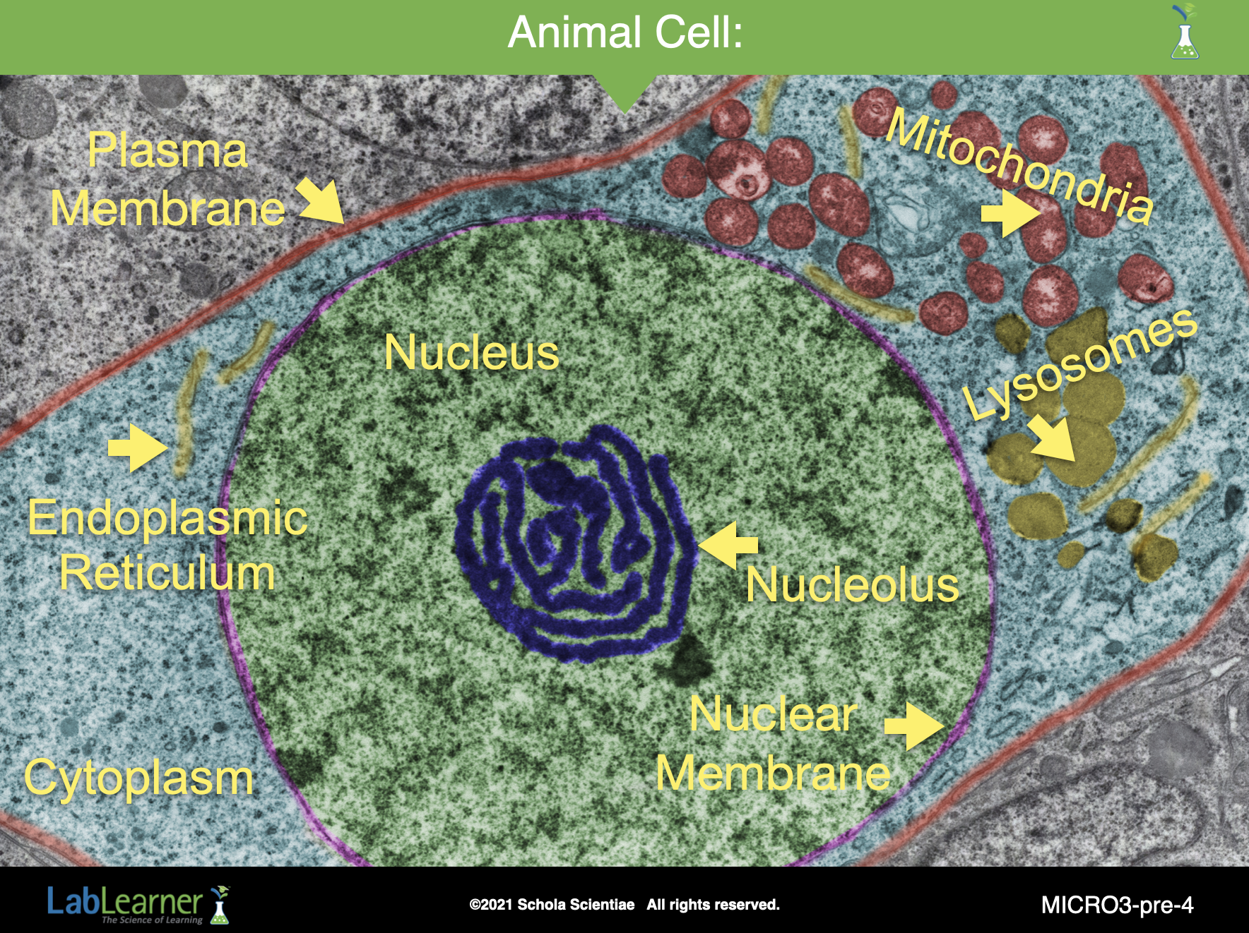
This artificially colored slide shows an electron micrograph (photograph) of a single animal cell in a tissue. This cell is separated from the rest of the similar cells in the surrounding tissue by a cell membrane (or plasma membrane), indicated by the red outline around it.
You can clearly see the large green nucleus surrounded by the nuclear membrane that separates it from the cytoplasm.
There are other structures in the cytoplasm that will be discussed shortly. These include mitochondria, lysosomes, endoplasmic reticulum, and many other membrane-covered organelles.
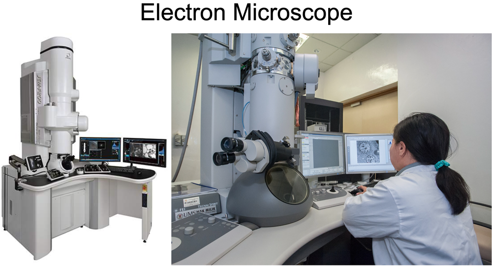
Electron Microscope: Electron microscopes use beams of electrons rather than light to magnify specimens to incredible magnifications. Images can be captured photographically at magnifications up to 50 million times! Electron microscopes are very expensive and very large scientific instruments. Look at the electron microscope pictured below. Most electron microscopes, like the one shown here, require their own special room with trained technicians and scientists to operate them.
______________________________________________
SLIDE MICRO3-pre-5
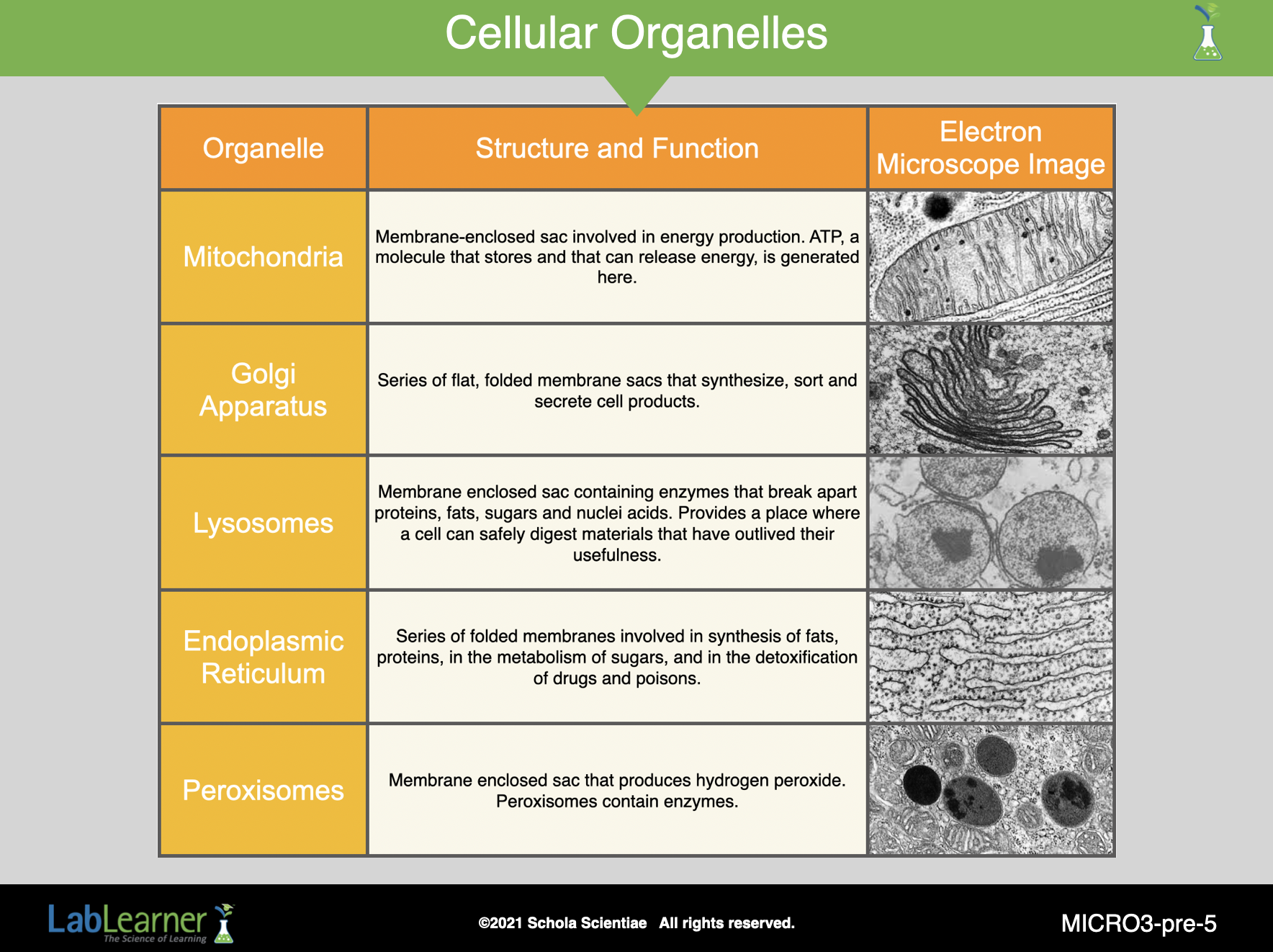
Organelles are membrane-bound (membrane-covered) structures found in the cytoplasm. Each organelle has a distinct function and together their combined functions allow cells to perform all of their important functions.
Mitochondria: membrane-enclosed sac involved in energy production. ATP, a molecule that stores and that can release energy, is generated here.
Golgi Apparatus: series of flat, folded membrane sacs that synthesize, sort, and secrete cell products.
Lysosomes: membrane-enclosed sac containing enzymes that break apart proteins, fats, sugars, and nucleic acids. Provides a place where a cell can safely digest materials that have outlived their usefulness.
Endoplasmic Reticulum: series of folded membranes involved in the synthesis of fats, proteins, in the metabolism of sugars, and in the detoxification of drugs and poisons.
Peroxisomes: membrane-enclosed sac that produces hydrogen peroxide. Peroxisomes contain enzymes.
These and other organelles coordinate in their functions to provide the cell with everything it needs to live, grow, and reproduce.
______________________________________________
SLIDE MICRO3-pre-6
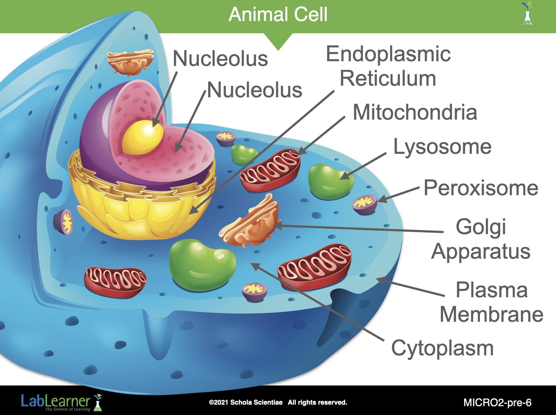
This slide is simply a three-dimensional model of an animal cell showing the nucleus and the cytoplasmic organelles that we discussed in the previous slide.
______________________________________________
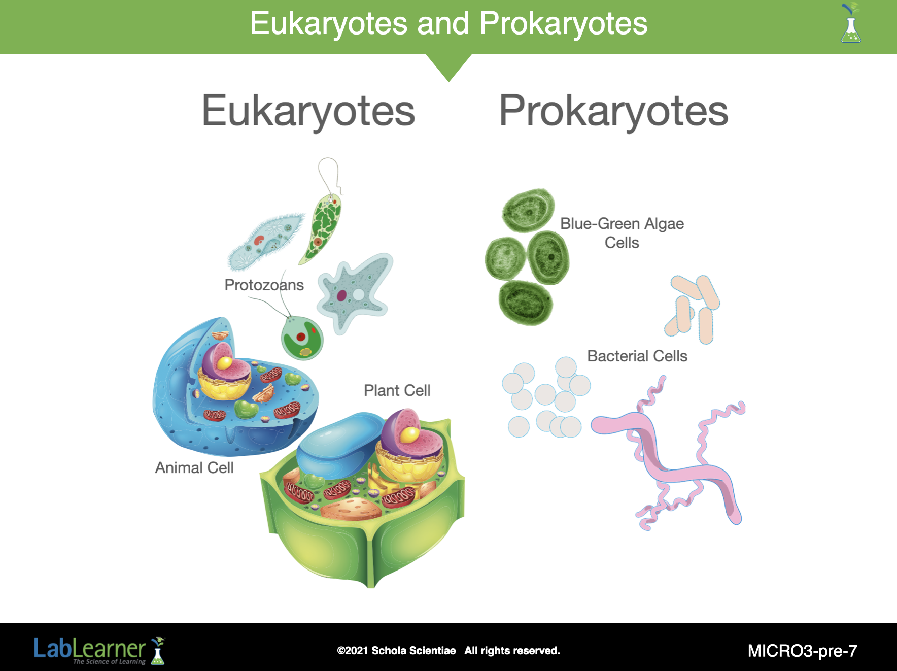
SLIDE MICRO3-pre-7
This slide shows a major division in life on Earth, eukaryotic and prokaryotic organisms.
Eukaryotes:
- Notice that, while some eukaryotes are single cells, like the protozoans, eukaryotes also include multicellular plants and animals. You are a eukaryote (“you-karyote”), for example.
- Every eukaryotic cell, both single-celled and multicellular organisms, and both plants and animals have a nucleus that is surrounded by a membrane, the nuclear membrane. In fact, the root karyon is Greek and refers to the nucleus.
- Eukaryotic organisms are also much larger than prokaryotic cells.
Prokaryotes:
- All prokaryotes are unicellular.
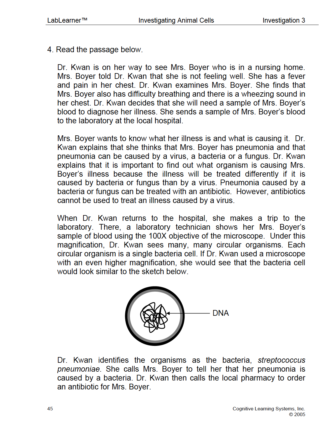
- Although prokaryotes have DNA, it is not surrounded by a nuclear membrane to separate it from the rest of the cytoplasm. Thus, we say that prokaryotes have no nucleus.
- Prokaryotes appeared on Earth way before eukaryotic organisms.
- Prokaryotic cells are much smaller than eukaryotic cells.
- Unlike eukaryotes, prokaryotes do not have organelles (mitochondria, lysosomes, etc.).
______________________________________________
PRELAB EXERCISE
In Investigation Three, students will use the compound microscope to view an animal cell taken from a living organism- themselves. The activity below has been included to help students understand what is meant by the term cell, as this may be students’ first exposure to the concept of cells.
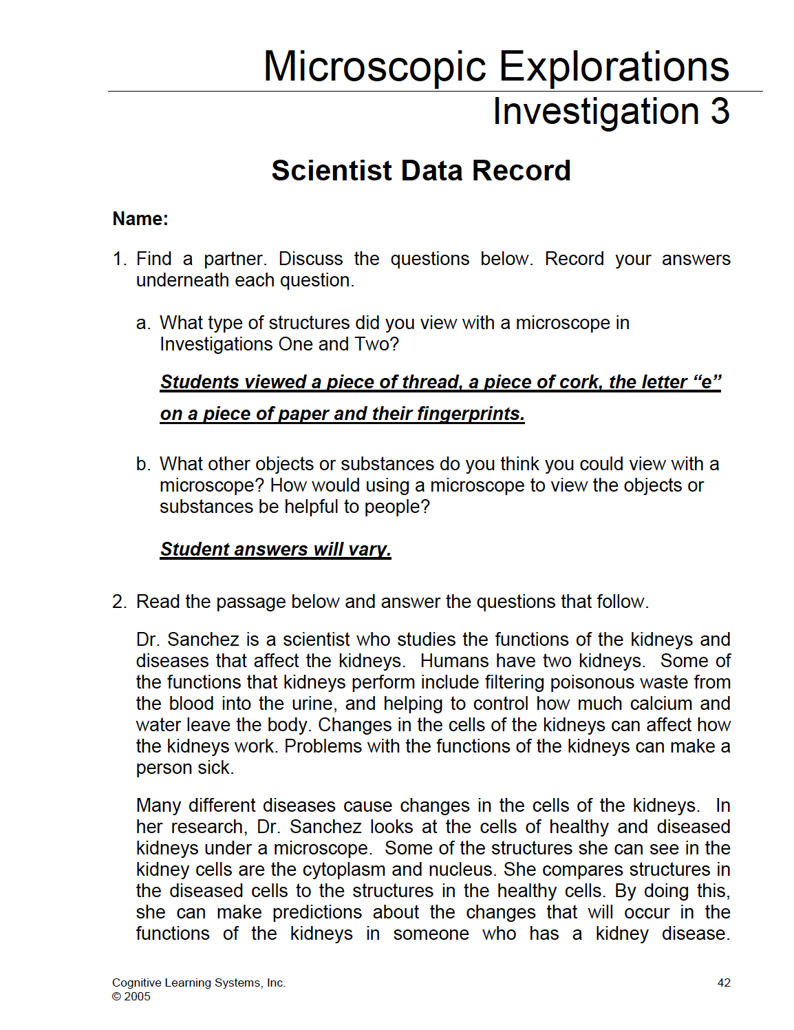
What type of structures did you view with a microscope in Investigations One and Two? Student answers will vary.
What other objects or substances do you think you could view with a microscope? How would using a microscope to view the objects or substances be helpful to people? Student answers will vary.
1. Ask students to find a partner and discuss the question. Students should then record their ideas in Problem 1 of their Scientist Data Record. Provide an opportunity for students to share their ideas with other members of the class.
2. After students have shared their ideas, direct them to Problem 2 of their Scientist Data Record and either read the passage aloud or ask them to read it independently.
KEYS: PRELAB EXERCISE

WATCH IT
Play the following Student Video in preparation for the lab. Discuss as necessary to answer student questions.