Teacher Portal:
Microscopic Explorations
Investigation 2
Microscopic Explorations: Investigation 2
Prior to jumping into the use of microscopes to study cells, we wish to take a few moments simply considering the size of things. Students appreciate that things come in many different sizes. However, the range in size is almost beyond human comprehension. From the incomprehensibly small to the incomprehensively large, things in our world come in every size. The graphic below fits a single human on a scale of objects ranging from the super small subatomic particles on the left to the entire Universe on the right.
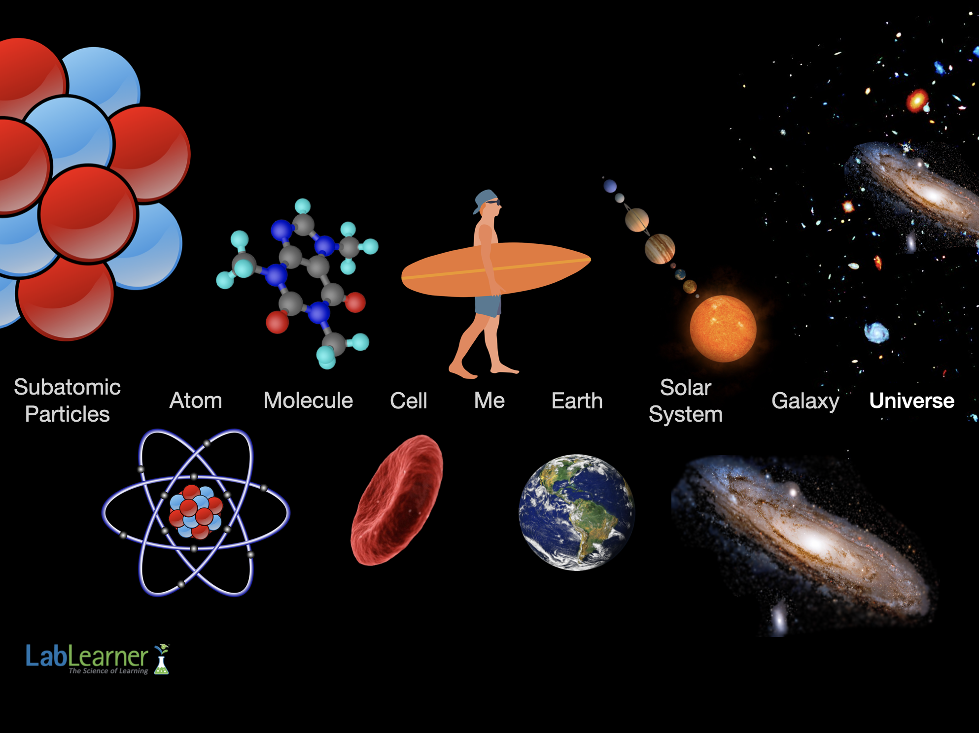
To study the objects to the right of the human shown in the above illustration, we can either use our unassisted eyes or better yet, a telescope. In many ways, modern compound microscopes and telescopes are closely related types of scientific instruments. Both use two or more lenses and direct light through an enclosed tube. To observe the objects to the left of the human in the above illustration, we must use a microscope. Our unassisted eyes cannot see a single cell.
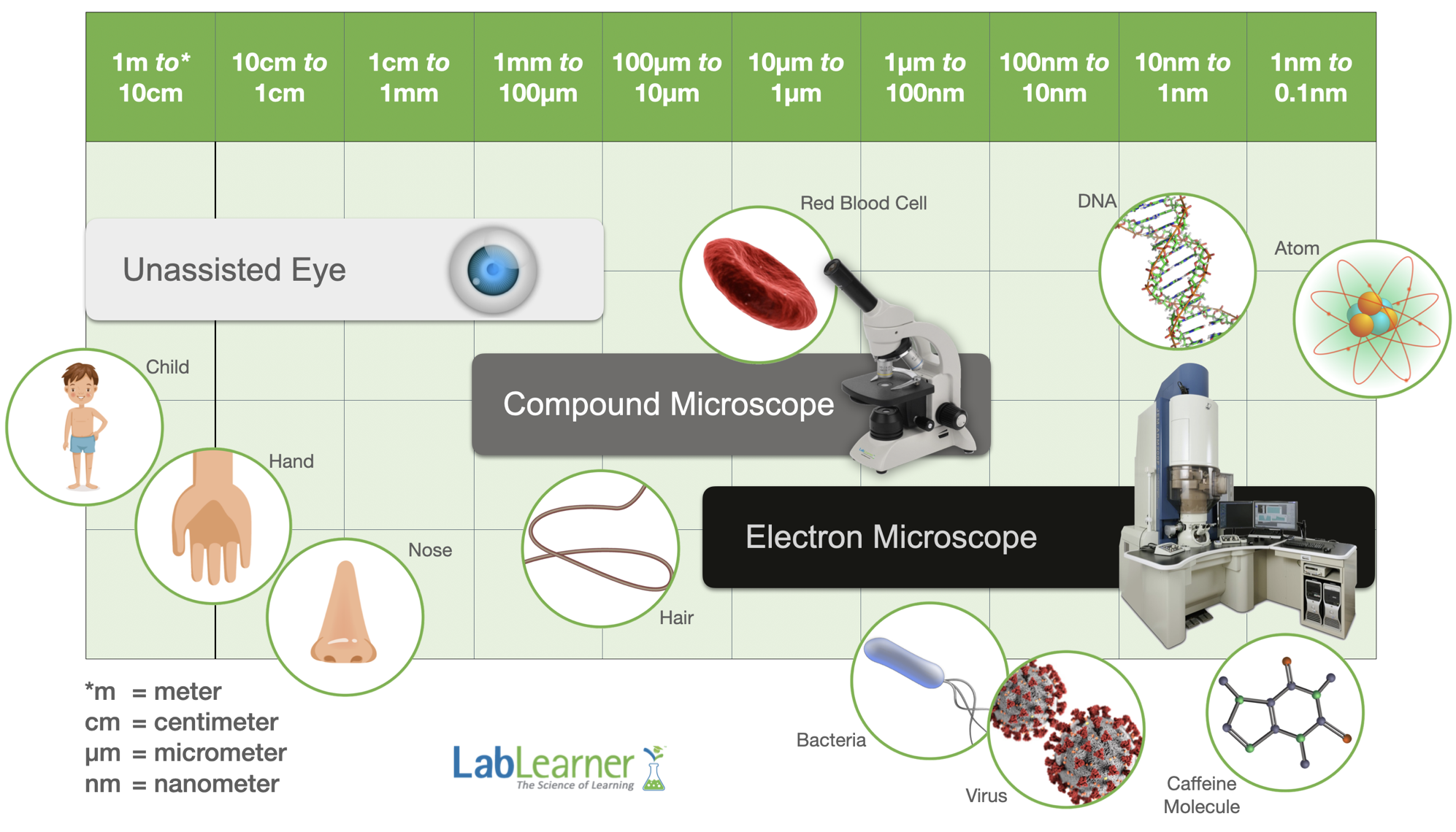
There are various types of microscopes. Two major types are compound microscopes, similar to the ones used in LabLearne, and electron microscopes. Electron microscopes are very large and expensive scientific instruments. An entire room often houses a single electron microscope. The illustration below once again depicts a range of microscopic objects and the instruments (unassisted eye, compound microscope, and electron microscope) used to view them.
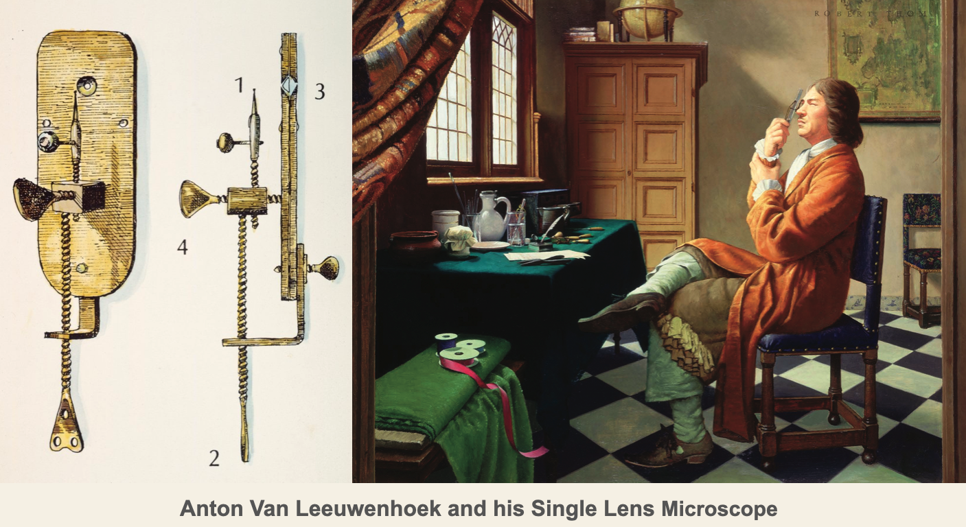
Investigation Two moves students from working with handheld lenses to using the lenses in the compound microscope. The compound microscope has a long history in the scientific world. Although much is publicized about the microscopes made and used by Anton Van Leeuwenhoek, this 17th-century biologist is not the originator of the compound microscope. Van Leeuwenhoek’s microscopes were simple microscopes in that they used a single convex lens for viewing specimens. In contrast, the compound microscope utilizes more than one lens. However, the inventor of the compound microscope is a title that has been given to more than one individual in the scientific community. Some credit Galileo Galilei and the compound microscope he developed in 1609 that used a convex and concave lens. Others point to Christiaan Huygens, a Dutchman, who developed a two-lens system in the early 1600s.
Later compound microscopes such as those used by Robert Hooke in the 17th century contained one convex lens in the objective and one in the eyepiece and served as the predecessor to the modern compound microscope. Modern compound microscopes such as the one students will use in this investigation contain several convex 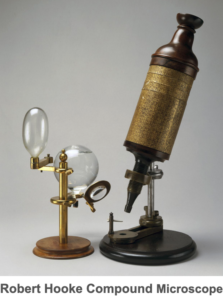 lenses of varying magnification arranged on a rotating nose piece and one convex lens in the eyepiece. Although only one objective is used at a time, the rotating arrangement of lenses allows the user to choose the level of magnification used for observation of a specimen.
In Investigation Two students continue their exploration of microscopy, lenses, and refraction. Students prepare a specimen for microscopic analysis and analyze prepared slides of cork, newsprint, and thread with the compound microscope. In addition to the concepts learned during Investigation One such as the ability of lenses to maintain the proportions of an object when producing its image, students’ experiments will focus on several new concepts. These include the calculation of the power of magnification, the relationship between magnification, resolution, and field of view, and the relationship between the structure and function of objects and substances. During their investigation, students will use not only the 4X (low power) objective of the microscope but also the 10X (medium-power) and 40X (high-power) objectives. As they observe specimens with each objective, they will be asked to determine the power of magnification that is observed by the human eye. In the compound microscope, the total power of magnification observed is the product of the magnification of the objective used and the magnification of the eyepiece.
Total power of magnification = objective magnification X eyepiece magnification. The magnification of the convex lens in the eyepiece of the compound microscope students will use is 10X. This means that if an object were  viewed solely through the eyepiece lens, it would appear ten times larger than it is. The power of magnification of the low, medium, and high power objectives are denoted on the objectives. The low power objective is labeled 4X, the medium power objective 10X, and the high power objective 40X. Therefore if a student observes a specimen using the 40X objective, the total power of magnification he or she will observe in the image would be 40X multiplied by 10X, or 400X. The image would appear 400 times larger than it actually is.
![]() In addition to their observance of the power of magnification, students’ attention will be drawn to what scientists term the field of view. The term field of view in microscopy refers to the portion of the specimen’s image that is observed. As students compare specimens under the three objectives they will discover a principle of lenses and microscopy: the higher or more powerful the magnification, the greater the resolution of the image, but the smaller the field of view. Thus, increases in magnification come at a cost: more detail of an object can be seen but less of the object can be seen at any one time. Although seemingly simple, understanding and utilizing this principle is important for a thorough analysis of a microscopic specimen. Observation of the specimen with low resolution under a large field of view often provides information about the overall structure of a specimen whereas observations at higher levels of magnification and resolution provide information about the smaller details of the specimen’s composition. During Investigation Two students will experiment with the use of different objectives, magnifications, fields of view, and resolution. In doing so, they will become more skilled at discerning both the structure and function of the specimens they investigate.
In addition to their observance of the power of magnification, students’ attention will be drawn to what scientists term the field of view. The term field of view in microscopy refers to the portion of the specimen’s image that is observed. As students compare specimens under the three objectives they will discover a principle of lenses and microscopy: the higher or more powerful the magnification, the greater the resolution of the image, but the smaller the field of view. Thus, increases in magnification come at a cost: more detail of an object can be seen but less of the object can be seen at any one time. Although seemingly simple, understanding and utilizing this principle is important for a thorough analysis of a microscopic specimen. Observation of the specimen with low resolution under a large field of view often provides information about the overall structure of a specimen whereas observations at higher levels of magnification and resolution provide information about the smaller details of the specimen’s composition. During Investigation Two students will experiment with the use of different objectives, magnifications, fields of view, and resolution. In doing so, they will become more skilled at discerning both the structure and function of the specimens they investigate.
Microscopic Explorations: Investigation 2 - Mathematics Concepts
Prelab
• comparing (non)measurable characteristics
• data table
• predicting/verifying size, shape, form
• greater than/less than/equal to
• least to greatest
Lab
• (in)direct relationships
• multiplication
• comparing (non)measurable characteristics
• length in cm
• volume in mL
• data table
Postlab
• data analysis
• multiplication/division
• comparing (non)measurable characteristics
• drawing to scale
• proportions/ratio
• least to greatest
• (in)direct relationships
Microscopic Explorations: Investigation 2 - Procedural Tools

Microscopic Explorations: Investigation 2 - Cognitive Tools
Microscopic Explorations:
Investigation 2 Quiz

