Teacher Portal:
Microscopic Explorations
Microscopic Exploration
 During this CELL, students will conduct Investigations focused on both understanding how the compound microscope works and utilizing the principles upon which its function is based in order to study microscopic structures of various specimens. Through their explorations, they will come to a better understanding of the field of microscopy and will discover how microscopy can be used to understand the structure and function of living and non-living things.
Students will investigate the effects of refraction of light by double convex and double concave lenses, examining the image produced by each type of lens. As students compare these images to the original object they will come to realize that changes in resolution can accompany refraction. By collecting quantitative data, students will also determine that the proportions of an object are maintained in the images produced by these lenses. Students’ investigations with lenses will serve as prior knowledge for understanding why the microscope can produce images of objects and structures that cannot be seen with the unassisted eye as well as for understanding changes in the field of view and resolution that accompany the magnification produced by the microscope. Students will then have the opportunity to utilize this knowledge as they prepare and analyze specimens for microscopic analysis. By manipulating the microscope to provide changes in resolution and field of view students will observe plant and animal cells and tissues as well as the organelles contained within them. Students will then have the opportunity to apply their knowledge from this CELL, by using microscopic analysis to determine the identity of unknown specimens from a mock crime scene. By the conclusion of this CELL, students should realize that microscopy is a field that utilizes the principles of light and physics to discover the structures and functions of the biological and physical world.
Through experiments, observation, microscopic analysis, and participation in class discussion, students will begin to comprehend that what they see with their unassisted eyes is only one part of the world around them. This CELL will promote students’ awareness of the microscopic structure of living and non-living things they see or experience on a daily basis, building continuity between laboratory investigations and real-world applications and encouraging independent investigation.
___________________________________________________________________
Investigation 1: Learning About Lenses
In Investigation One students will explore the effect of the refraction of light through lenses. By observing objects with hand-held convex and concave lenses and the convex lenses in a compound microscope, they will discover that refraction of light through lenses produces images that differ in resolution from the object. As students compare the images they will learn that the changes in resolution produced by convex and concave lenses are different. Compared to the object, images produced by convex lenses have increased resolution while those from concave lenses are decreased in resolution. Students’ observations will also enable them to realize that when convex and concave lenses are used, the proportions of the object are maintained in the image.
Investigation 1: Teacher’s Video (10:04)
Investigation 1: Student’s Video (16:00)
___________________________________________________________________
Investigation 2: Investigating Specimens
In Investigation Two, students will build on their experience with lenses as they use the three objectives of the compound microscope to study specimens. Their observation of the specimens will enable them to discover that refraction of light through lenses not only produces changes in magnification and resolution but also in the field of view. As a part of their analysis, students will calculate the total power of magnification produced when specimens are viewed with each objective. In addition, students will be introduced to specimen preparation techniques as they prepare their fingerprints for analysis under the microscope.
Investigation 2: Teacher’s Video (13:01)
Investigation 2: Student’s Video (16:46)
___________________________________________________________________
Investigation 3: Investigating Animal Cells
During Investigation Three, students will be introduced to the concept of a cell and to examples of eukaryotic and prokaryotic cells. Students will learn the procedure for a wet mount preparation of a specimen as they prepare a sample of their own cheek cells for analysis. Through observation of their cheek cells, students will discover that a cell is composed of smaller parts called organelles and that the structure of a cell and its organelles relate to its functions.
Investigation 3: Teacher’s Video (11:01)
Investigation 3: Student’s Video (4:18)
___________________________________________________________________
Investigation 4: Investigating Plant Cells
Investigation Four introduces students to examples of plant cells. Students will observe specimens of an Elodea leaf and an onion bulb. During their investigation, they will discover that plant cells have a cell wall, an organelle not present in animal cells. Through their observations, students will also realize that chloroplasts, organelles absent in animal cells, are present in some but not all plant cells and that this difference is directly related to the ability of plants to carry out photosynthesis.
Investigation 4: Teacher’s Video (16:29)
Investigation 4: Teacher’s Video: Leaves & Pigments (2:24)
Investigation 4: Student’s Video (13:13)
Click on the image below to open Investigation 4 CAP
___________________________________________________________________
Investigation 5: Investigating Tissues
In Investigation Five, students will shift the focus of their microscopic exploration from cells to tissues and organs. By observing specimens of the leaves and stems of plants and blood and the colon of the human body, students will come to realize that cells combine within organisms to form larger structures. Through comparison of the cell shapes, sizes, and arrangement within the four specimens students will develop a deeper understanding of how the function of a tissue or organ relates to its structure.
Investigation 5: Teacher’s Video (9:02)
Investigation 5: Student’s Video (10:59)
___________________________________________________________________
Investigation 6: Performance Assessment
Investigation Six consists of a performance assessment designed to evaluate students’ understanding of the science concepts addressed in the first five Investigations of the Core Experience Learning Lab. During this exploration, students will use the knowledge they have gained about lenses, resolution, microscopic analysis, specimen preparation, and cells to identify three unknown samples from a crime scene.
Investigation 6: Performance Assessment – Teachers Only (14:21)


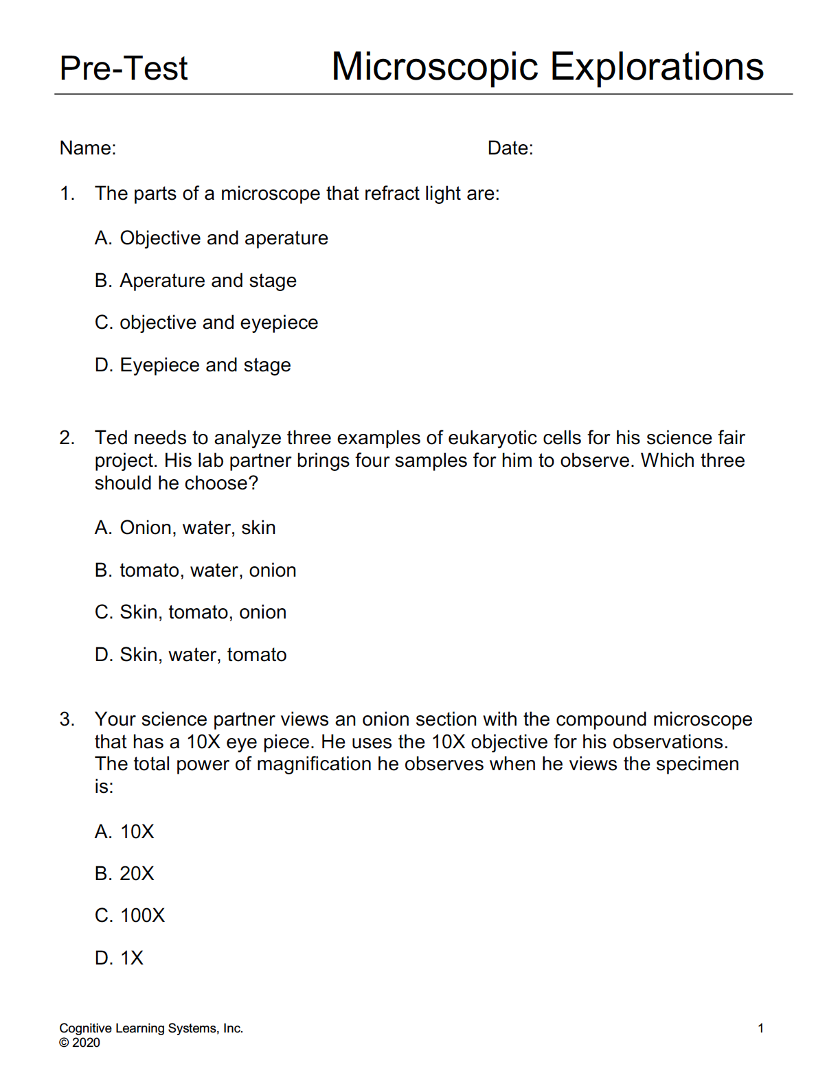
Pre-Tests and Post-Tests
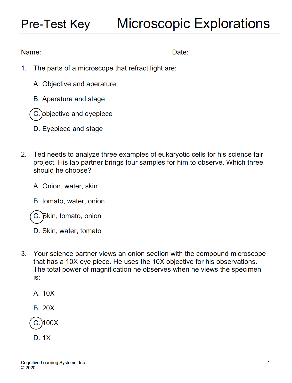
Pre-Test Key
Includes NGSS correlations
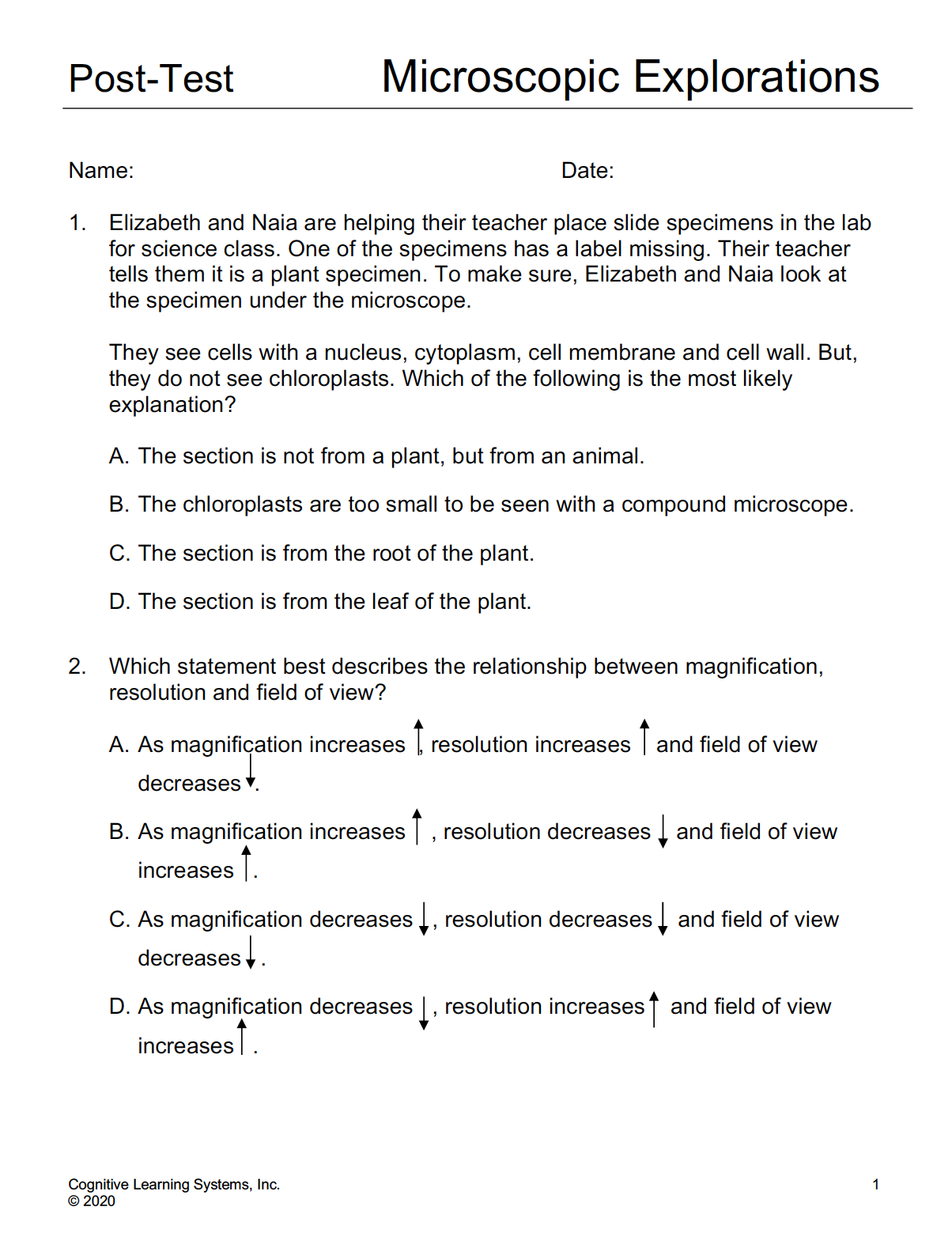
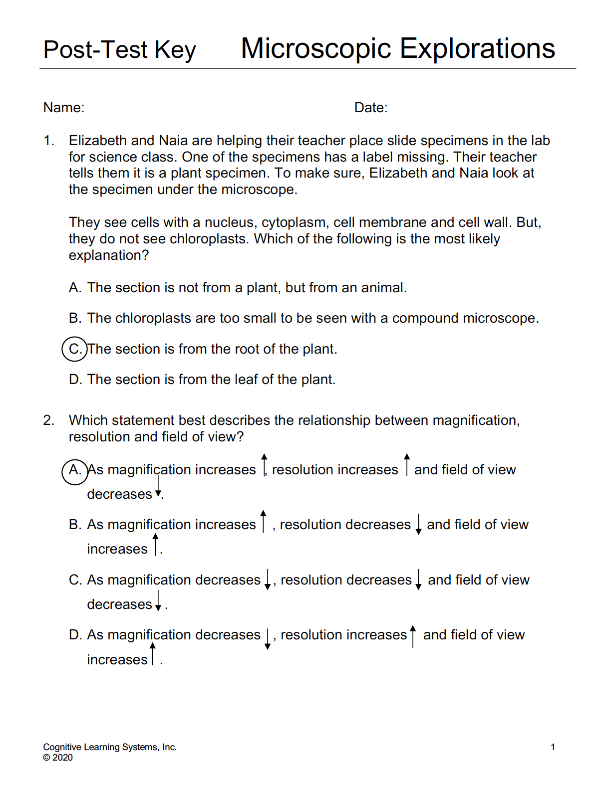
Post-Test Key
Includes NGSS correlations