Teacher Portal:
Examining Exercise
Investigation 1 – Lab
 BE PREPARED
Supplies and Equipment:
Class Materials:
- 1 human torso
- 1 human skeleton
Pair Materials:
- 1 spring scale
- 1 stopwatch
- 1 stethoscope
- 2 alcohol swabs
- 1 calculator
Individual Materials:
- 1 Student Data Record
Teacher Preparation:
1. Position the human torso and skeleton at the front of the lab.
2. Organize the required materials at a distribution point.
3. Separate the class into pairs.
Instruction:
1. Direct each student pair to obtain the following necessary materials from the distribution point: one (1) spring scale, one (1) stopwatch, one (1) stethoscope, two (2) alcohol swabs, and one (1) calculator.
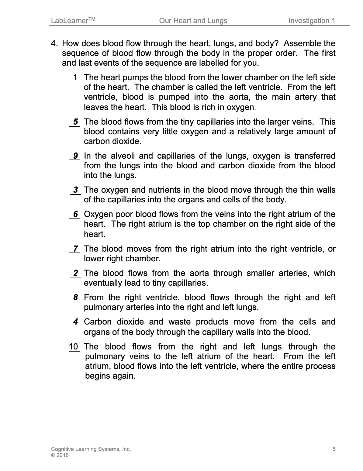
2. Review the flow of blood through the cardiovascular and respiratory system with students using the human torso and flipchart visual aids. After the review, encourage students to describe the sequence of the flow of blood through the heart, lungs, and body by completing Problem 4 in their Student Data Record.
GET FOCUSED
Investigation One introduces students to pulse and heart rate.
INVESTIGATE
1. Students will observe the human torso, flipchart, and skeleton to explore the respiratory system. In addition, students will use a spring scale to model and analyze the process of respiration. The use of the spring scale will help increase students’ understanding of how the lungs, rib cage, and diaphragm work together during exhalation and inhalation. The passages that follow are provided to assist your direction of the students through this part of the Investigation. Encourage students to complete Problem 5 in the Student Data Record as the activity is completed.
a. Explain that the diaphragm is a muscle located below the lungs and above the stomach and liver. It divides the thoracic (chest) and abdominal cavities.
Show students the location of the diaphragm on the human torso model and flipchart. The diaphragm is the muscle located on the bottom of each lung on the torso.
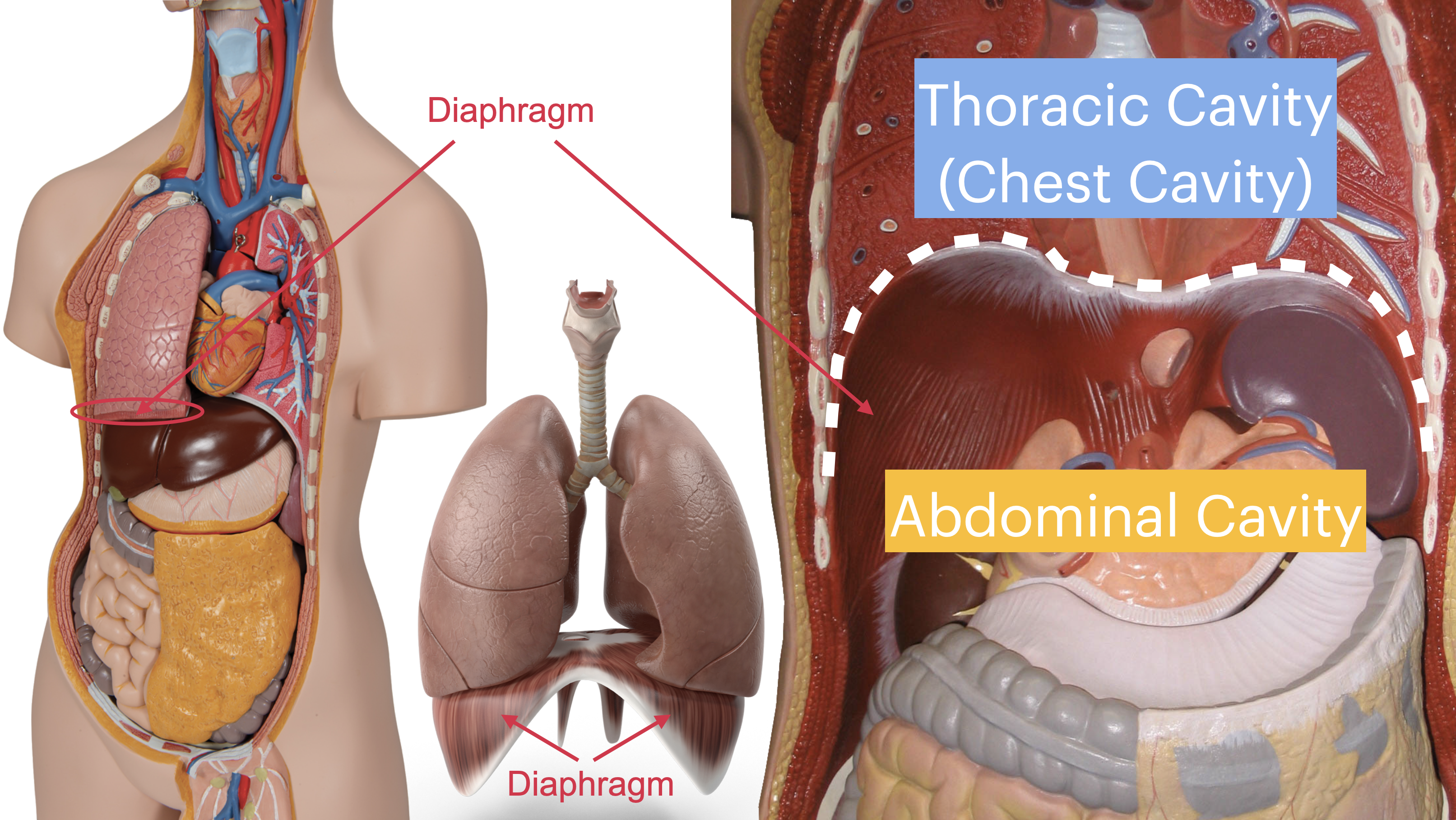
Explain that the diaphragm is also attached to parts of the skeletal system.
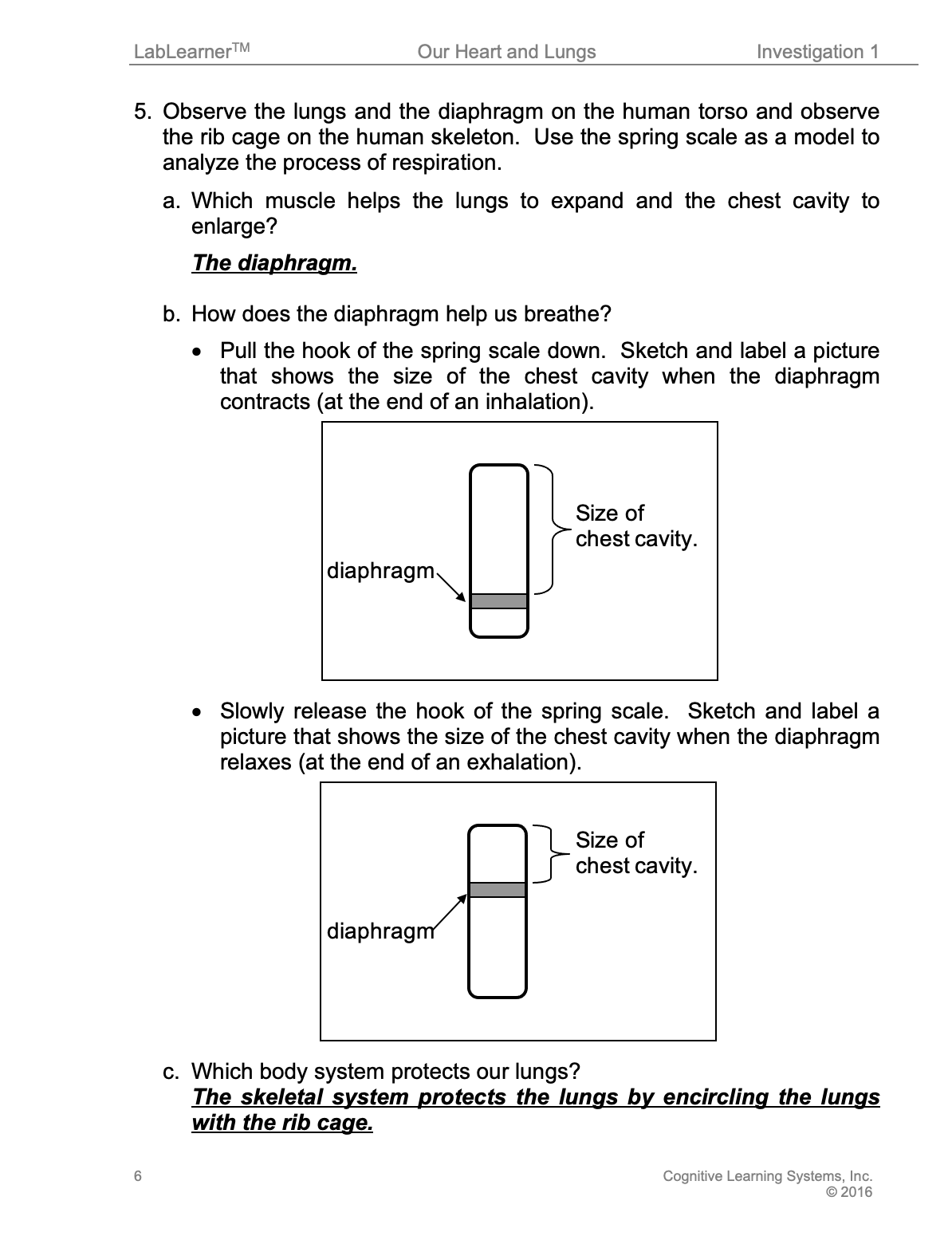
b. Explain that a spring scale can be used as a model of the chest cavity, or area including the ribs, the space inside the ribs, and the lungs. The platform of the spring scale represents the diaphragm.
c. Demonstrate the process of inhalation using the spring scale.
-
- As the diaphragm contracts and is pulled down, the chest cavity is enlarged.
- This allows the lungs to expand and bring air from the atmosphere into the body.
- Direct students to pull on the hook of the spring scale, which lowers the platform and increases the space above it.
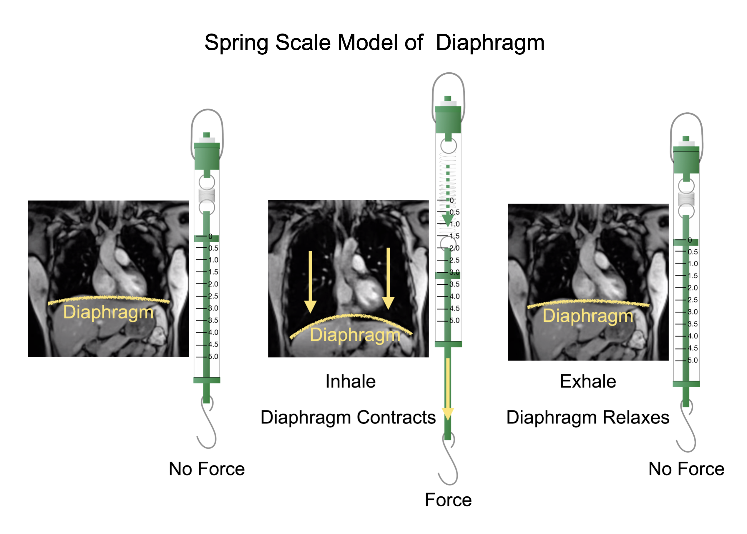
d. Demonstrate the process of exhalation using the spring scale.
-
- The diaphragm returns to its original position, and the chest cavity becomes smaller.
- The lungs are squeezed and air is forced out into the atmosphere.
- Direct students to slowly release the hook of the spring scale, which allows the platform to rise and decreases the space above it.
_________________________________________
2. During this part of the Investigation, students will observe the locations on their bodies where they can see their cardiovascular system, feel their heartbeat and determine their heart rate. The activities have been designed to draw students’ attention to the observable presence of the cardiovascular system on their body and how its structure within the body allows us to monitor its function.
This part of the Investigation has also been designed as a way to introduce students to the method of taking a pulse and as a way to review the calculation of heart rate. Students will be asked the following question in order to direct their attention to the way in which the structure of the cardiovascular system (arteries and veins present close to the skin) permits the determination of its function (heart rate).
How do you know your cardiovascular system is working?
3. Discuss the structures and functions of the respiratory system with students.
a. Use the human torso and human body flipchart during the discussion. Remove both the left and right lungs and pass each around for students to observe.
b. Direct students to inhale and exhale deeply, describing what occurs to the chest, lungs, and air inside and outside the body during each step.
c. As you facilitate these activities encourage students to record their observations and conclusions in their Student Data Record.
_________________________________________
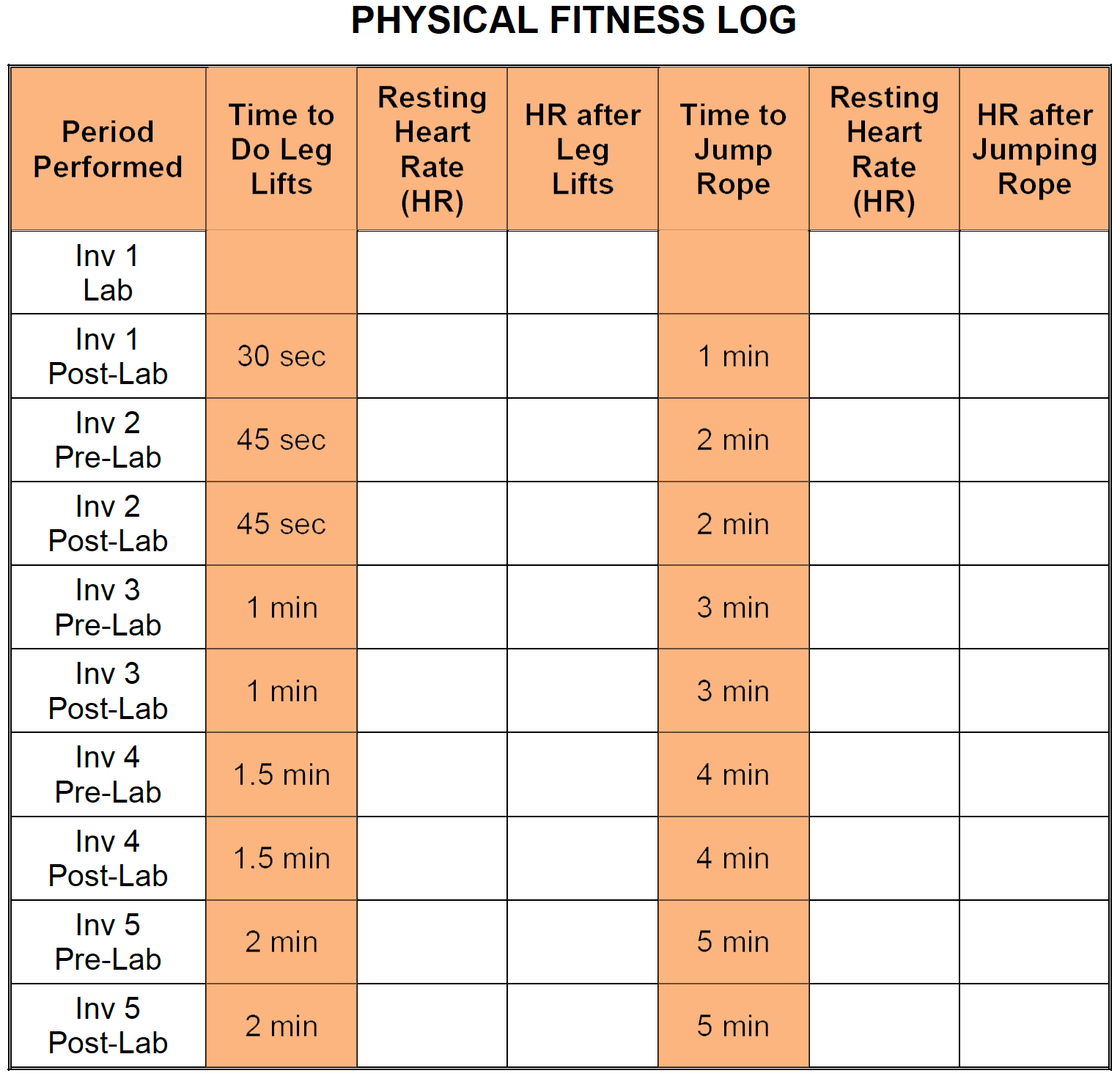
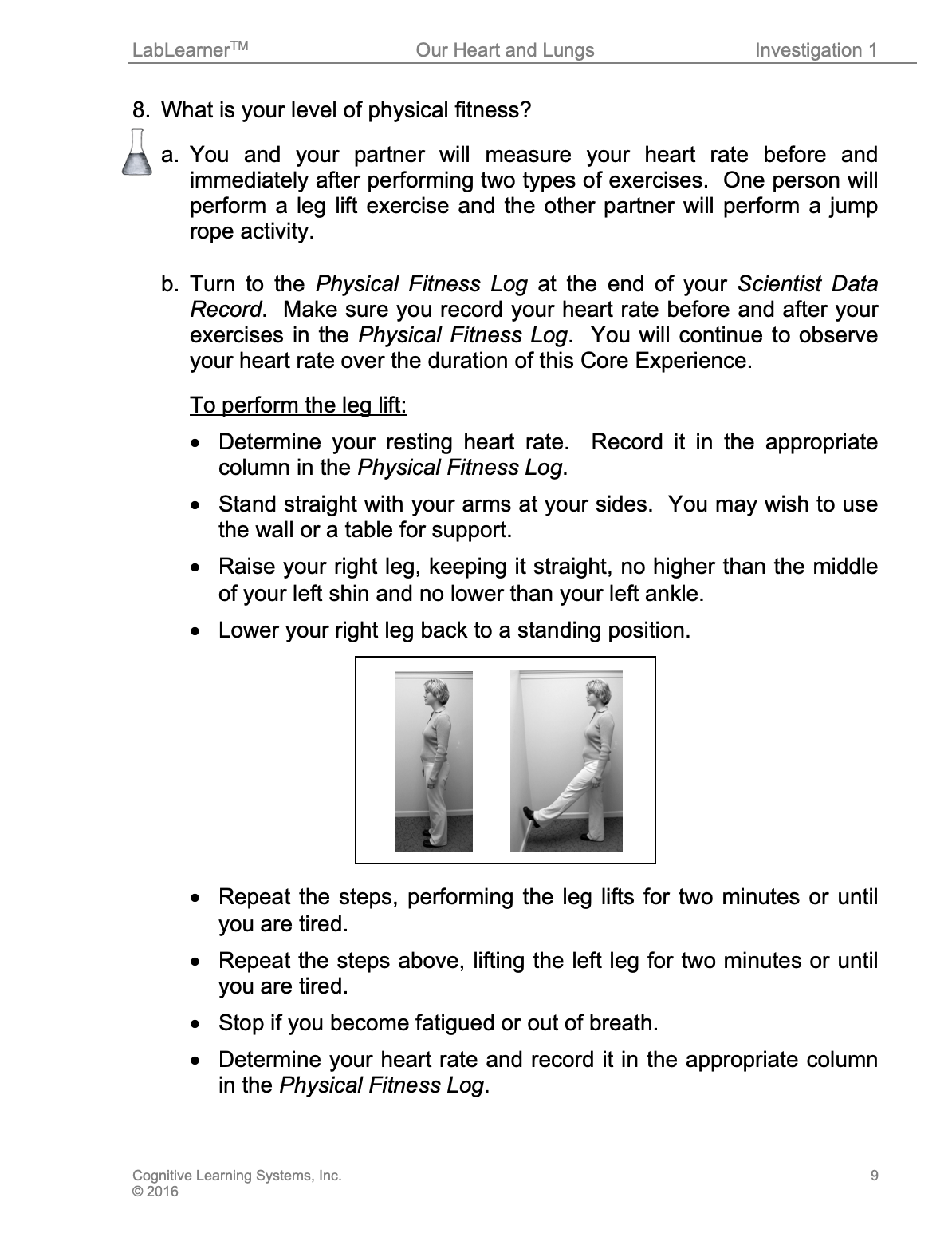
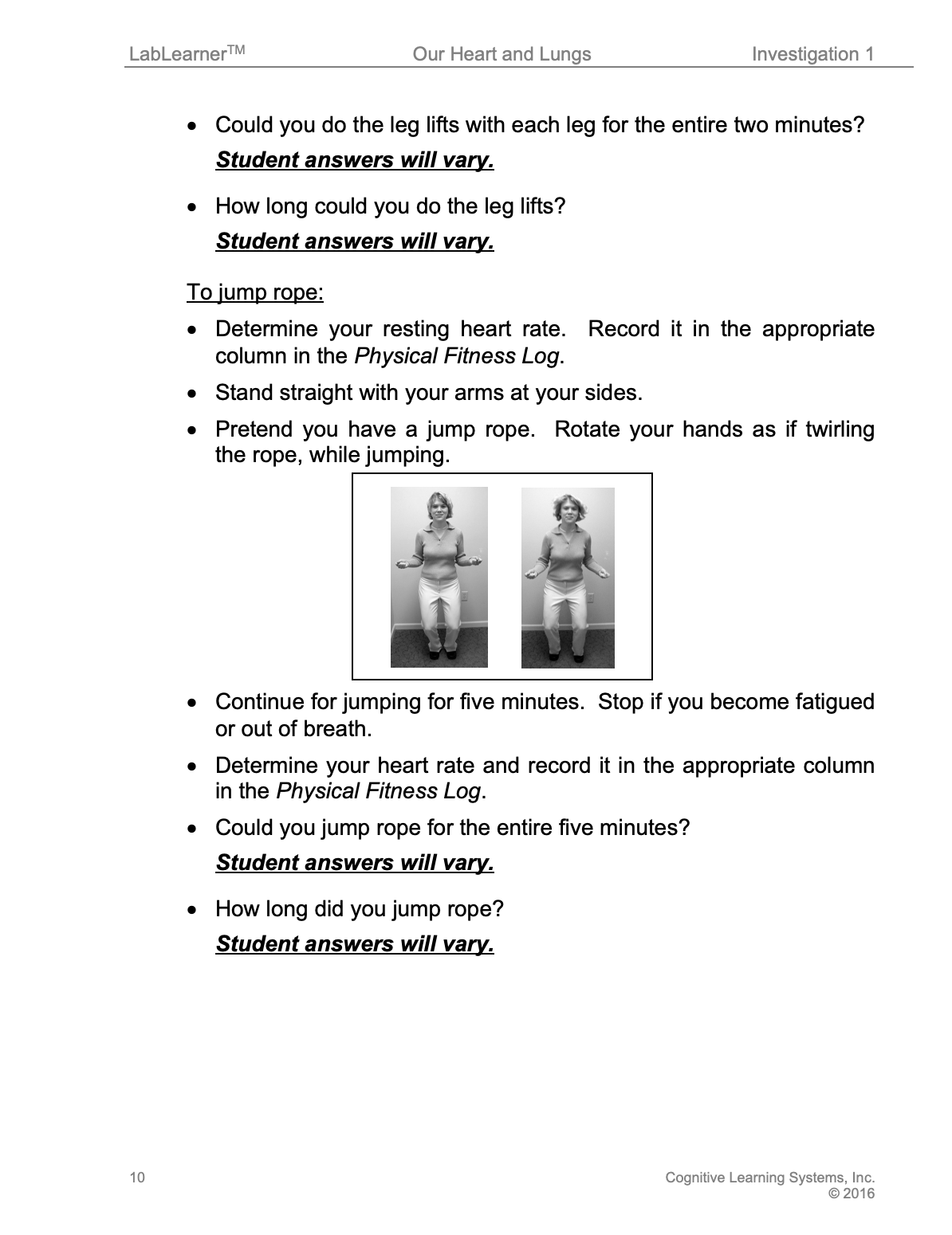
4. During this part of the Investigation, students will perform either leg lifts or participate in a jump rope-like activity. They will record their heart rate before and immediately after they perform their exercises in a Physical Fitness Log located in their Student Data Record. This is the first of several times that students will perform these activities. Students will perform these activities during each Pre-Lab and Post-Lab of the CELL as a way to illustrate the effects of regular exercise and training on heart rate and on cardiovascular and muscular endurance.
This ongoing experiment has been designed so that students are first challenged to perform leg lifts with each leg for two minutes and a jump rope activity for five minutes; a task most students should find challenging. Beginning with the Post-Lab in Investigation One, students participate in a “training” regime in which they perform the same activities for shorter periods of time. With each new Investigation, the training times become increasingly longer, thus promoting a slow but significant progression of cardiovascular and muscular endurance.
At the conclusion of the CELL, students will analyze the effect of both types of exercise on heart rate in addition to the effect (and benefits) that the regular performance of exercise (training) had on their cardiovascular and muscular endurance.
To begin their investigation, students will be asked the following question: What is your level of physical fitness?
KEYS

CLEAN UP
Let students know your expectations for clean-up. Ask them to clean up.