Teacher Portal:
Microscopic Exploration
Investigation 5 – PreLab
ASK WHY
Microscopes are one of the most important scientific instruments developed. In fact, in the medical field, microscopes are largely responsible for making modern medicine “modern”!
BRANCH OUT
Cell Biologists study how cells work. Cell Biologists are involved in important research in many different areas of biological sciences. They are employed at universities, hospitals, clinics, and industry.
PRINT IT
Use your browser to download a printable PDF as help during the slide presentation and to make additional notes. In your browser, go to File > Print and then choose to save as PDF.
MINDSET
This Investigation is designed to:
- provide students with an opportunity to view several samples of plant and animal specimens with the compound microscope.
- allow students to investigate how cells combine to form tissues.
- provide an opportunity for students to compare cells, tissues, and the arrangement of cells and tissues in different plant specimens, different animal specimens and between plant and animal specimens.
- encourage students to realize that although animals cells may contain some of the same organelles, there is variation in the shape of animal cells and in the arrangement of the cells in tissues
- encourage students to realize that although plant cells may contain some of the same organelles, there is variation in some of the organelles that are in cells, in the shape of plant cells and in the arrangement of the plant cells in tissues
- promote student understanding that there is a relationship between the structure of tissue, or organ and its function.
SCIENTIST’S GLOSSARY
1. Cell membrane: the outer layer of a cell. The cell membrane helps control what can enter and exit a cell.
2. Cell wall: the non-living structure that surrounds the cell membrane of most plant cells. The cell wall helps the plant cell keep its shape.
3. Chloroplast: organelle in plant cells where photosynthesis occurs.
4. Cross section: a piece of a specimen that is cut across the long axis of the specimen. A cross-section is the opposite of a longitudinal section.
5. Cytoplasm: the gel-like area of the cell between the nucleus and cell membrane where many of the functions of the cell occur.
6. Eukaryotic cell: a cell that contains a defined nucleus.
7. Longitudinal section: a piece of a specimen that is cut along the long axis of the specimen. A longitudinal section is the opposite of a cross-section.
8. Nucleus: small sac inside a cell that contains or contained DNA. One type of organelle in a cell.
9. Organ: One of the parts of the body that performs a function. Different tissues combine to form organs. The brain, heart, and stomach are examples of organs.
10. Organelle: a small structure inside a cell that performs a specific function.
11. Prokaryotic cell: a cell that does not contain a clearly defined nucleus.
12. Tissue: a collection of similar cells that form the structures of a plant or animal and work together to perform a function.
BE PREPARED
Watch the Investigation 5 Teacher Video below to prepare for the PreLab.
SET FOR SUCCESS
- Tell students that they are about to begin their study of tissues.
- Ask students to share the kinds of things they expect they might learn in these Investigations.
- Tell students they will again use the compound microscope, this time to examine plant and animal tissues.
Begin the PreLab Concept Slides to start students on their learning journey. Then watch the Pre-Lab Student Video as a class.
NOTE THAT THE PRELAB EXERCISE INVOLVING MODELING OF CROSS-SECTIONS AND LONGITUDINAL SECTIONS IS OMITTED FROM THIS PRELAB. THIS INVOLVES STUDENT SDR PAGES 76 (BEGINNING WITH PROBLEM 2) THROUGH 80.
NAVIGATE IT
Once the slide presentation is launched
- use your left and right arrows to advance or go back in the slide presentation, and
- hover your mouse over the left edge of the presentation to get a view of the thumbnails for all the slides so that you can quickly move anywhere in the presentation.
- Click HERE to launch the slide presentation for the CELL.
SHARE IT
SLIDE MICRO5-pre-1
In Investigation Five, we will discuss higher orders of organization above the level of individual cells. As we shall see, cells organize into tissues, which in turn can organize with different types of tissues to form organs and then organ systems, like the muscle system, or the nervous system, and so on.
______________________________________________
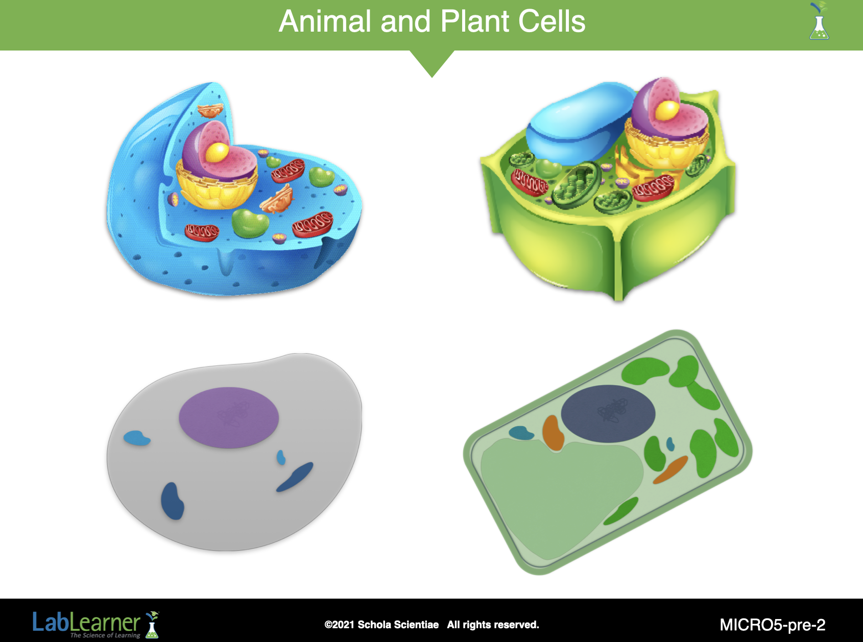
SLIDE MICRO5-pre-2
Cells are found in living organisms and contain many different structures. What are some structures that you would expect to find in a cell? Students should indicate that they would expect to find a cell membrane and cytoplasm in a cell. They may suggest that some but not all cells may also contain a nucleus.
How would you describe the size of most cells? Can cells be seen with the unassisted eye? Students should indicate that most cells are microscopic and cannot be viewed with the unassisted eye. A microscope is needed to view most cells. As an example, students may recall that their cheek cells were approximately 0.0015 cm in length.
Note: In general, the cells of most organisms are microscopic. However, there are some exceptions such as the giant squid axon and birds eggs which can be viewed with the unassisted eye.
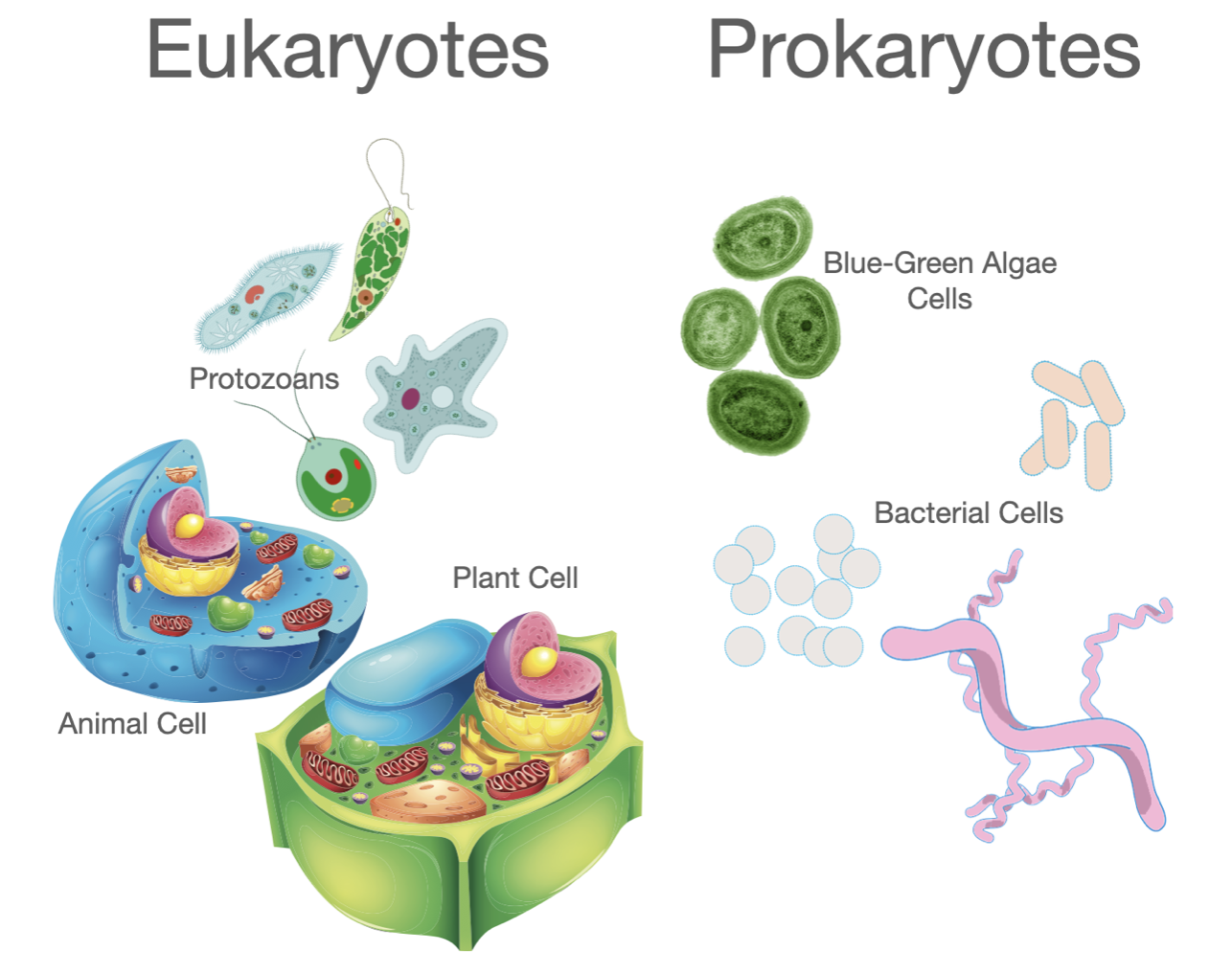
One way to classify cells is to categorize them as eukaryotic or prokaryotic. Describe the difference between a eukaryotic and prokaryotic cell. Students should indicate that a eukaryotic cell is one that contains a clearly defined nucleus, a nucleus that is separated by a membrane from the cytoplasm. A prokaryotic cell does not contain a clearly defined nucleus.
Do the parts of a cell have a function? What do the structures of the parts of a cell tell you about their functions? Students should suggest that the parts of a cell do have a function. Some of the functions of the different parts of a cell can be inferred from their structures. For example, the cell membrane surrounds a cell. It separates the inside from the outside of a cell. Therefore, it is likely that the cell membrane helps to control substances that can enter and exit a cell.
The organelles in a cell have functions. What would be examples of some functions of a cell? Students should indicate that cells have functions. For instance, cheek cells line the mouth, protecting the blood vessels and muscles of the mouth from food, enzymes, and bacteria; and the cells of an Elodea leaf perform photosynthesis, providing food for the plant.
Where would you expect to find cells? Can you give some examples of living organisms that have cells? Student answers may vary. Students should suggest that cells can be found in living organisms such as onion plants, Elodea plants, and humans.
______________________________________________
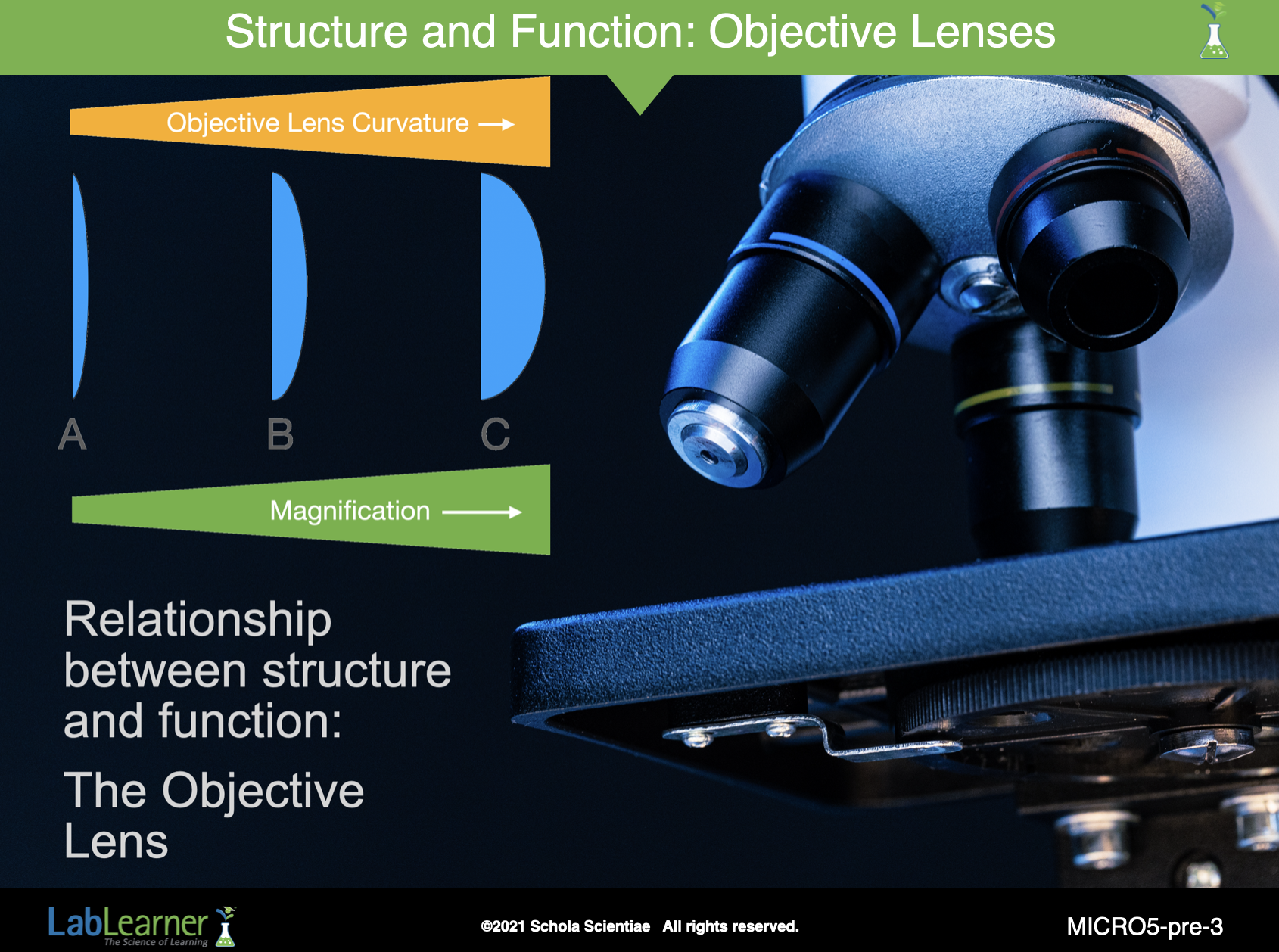
SLIDE MICRO5-pre-3
This is the first of three slides that focus on the relationship between structure and function. Students recently learned that the shape of a convex lens in an objective lens of a compound microscope determines how an image will look when viewed through it.
Increased curvature of the lens (structure) results in a greater magnification of the image (function).
______________________________________________
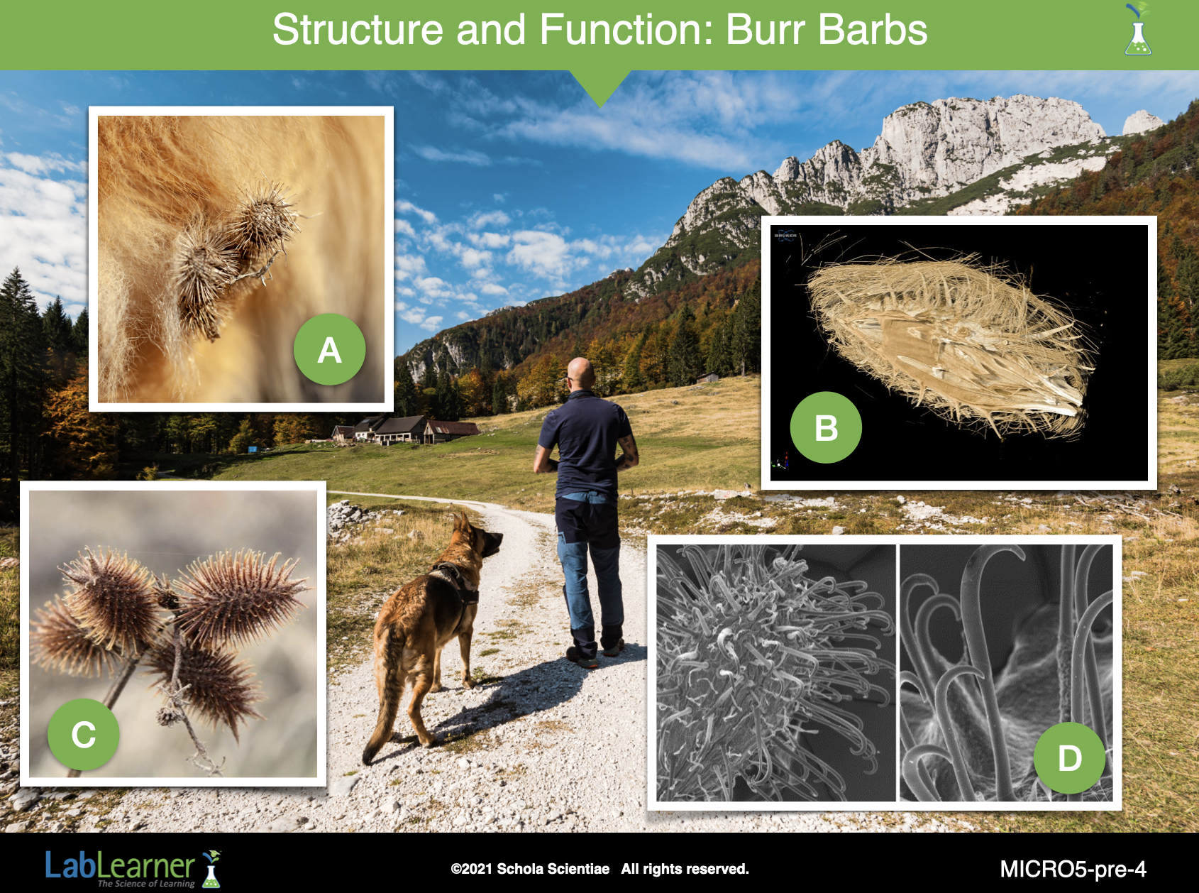
SLIDE MICRO5-pre-4
This slide and the next slide explain the relationship between structure and function in another example.
A Swiss engineer called George de Mestral was walking his dog in the Alps. When he returned home, he found that numerous plant seed burrs were tangled in his dog’s fur (A). He found that he could pull the burrs free from the dog but when examined under a hand lens, he saw that the burr was actually tangled in fur (B).
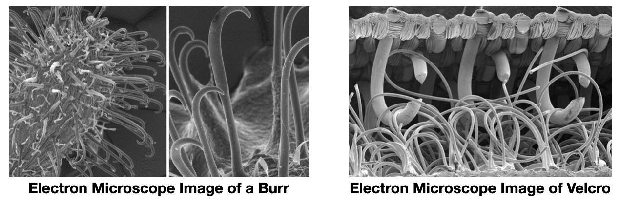
When burrs (C) are examined closely in a microscope (D), they are found to have tiny hooks on the end of their barbs. Thus, the microscopic structure of the burr helps to explain its function. In this case, the function of reversibly sticking to animal fur is to help the plant spread its seeds in the burr far from the plant so that the species can spread.
However, de Mestral was not finished. As an engineer, he thought that one surface reversibly binding to another might have useful applications as a fastener. And so, he used these ideas to invent…
______________________________________________
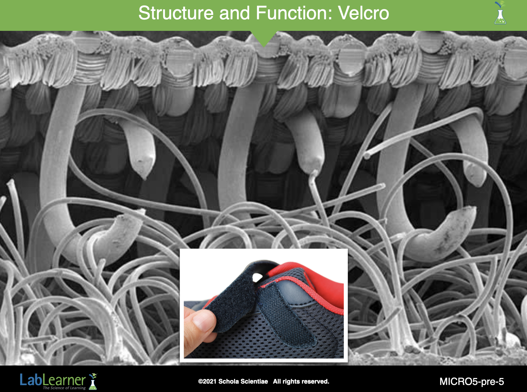
SLIDE MICRO5-pre-5
Velcro!
This slide shows a close-up electron microscope image of a small section of Velcro. Notice how the hook structures on the top surface interact with the fuzzy surface on the bottom. Further, notice how similar the Velcro microscopic image looks to the microscopic image of the burr on the previous slide.
______________________________________________
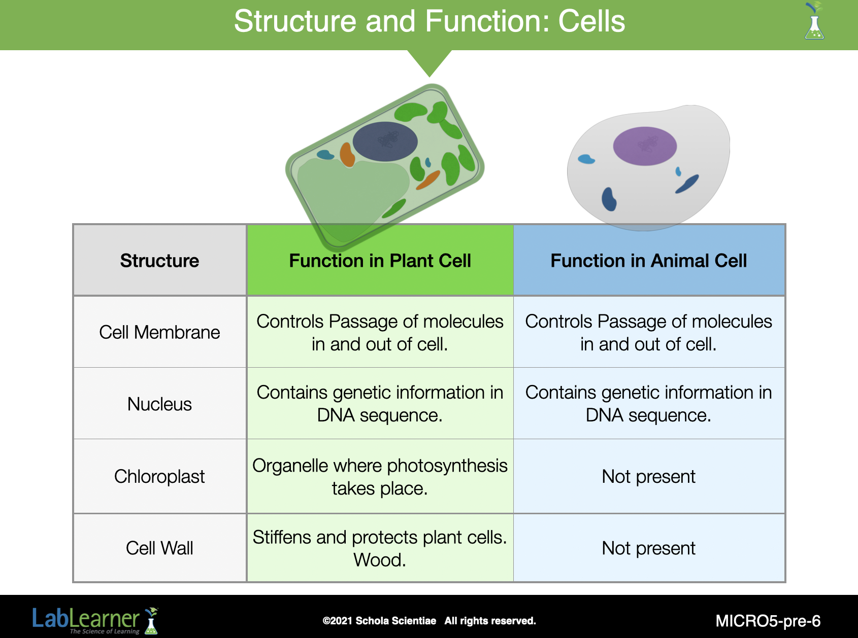
SLIDE MICRO5-pre-6
With this slide, we return to the subject of cells. Notice how we can extend a discussion of structure and function by comparing the structures in cells (organelles) to their functions.
Notice that the same structures that exist in both plant and animal cells perform similar functions (cell membrane and nucleus in this example).
Also notice that, if a specific organelle is missing from a cell (no chloroplasts in animal cells), that function is also missing from that cell (photosynthesis in this case).
______________________________________________
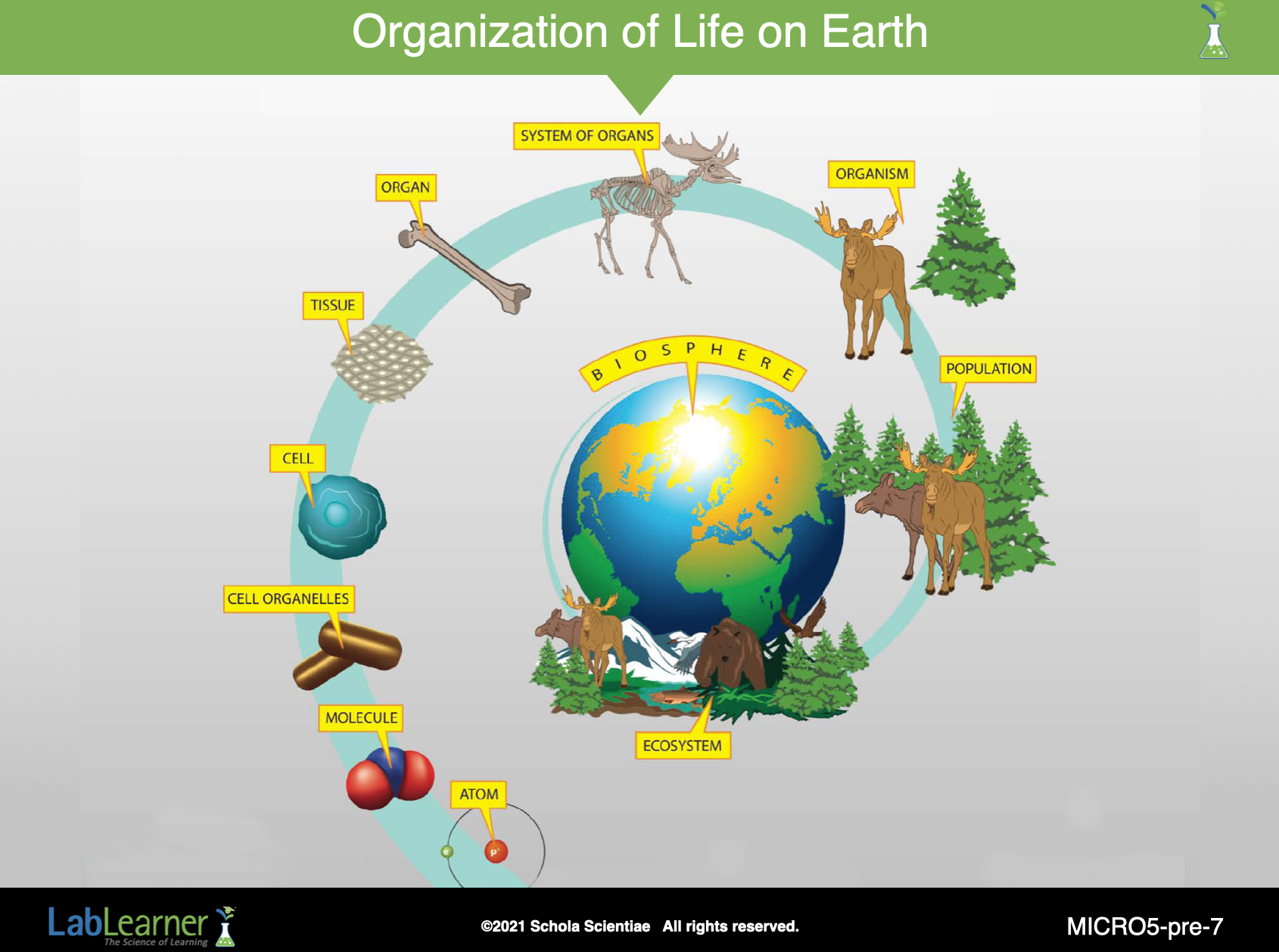
SLIDE MICRO5-pre-7
Before continuing further with the Investigation, assess students’ prior knowledge of the terms tissue, organ, and organism. If students are unfamiliar with these terms or are unsure of how to define these structures, direct them to locate the terms tissue and organ in their Scientist’s Glossary. Discuss examples of organisms such as humans, cats, trees, plants, reptiles, and birds.
Cell: the basic unit of organization of living organisms. Single-cell organisms live independently while other types of cells are organized into tissues.
Tissue: a collection of cells that form the structures of a plant or animal and work together to perform a function.
Organ: One of the parts of the body that performs a function. Different tissues combine to form organs. The brain, heart, and stomach are examples of organs.
Organism: an individual animal, plant, or single-celled life form.
______________________________________________
SLIDE MICRO5-pre-8
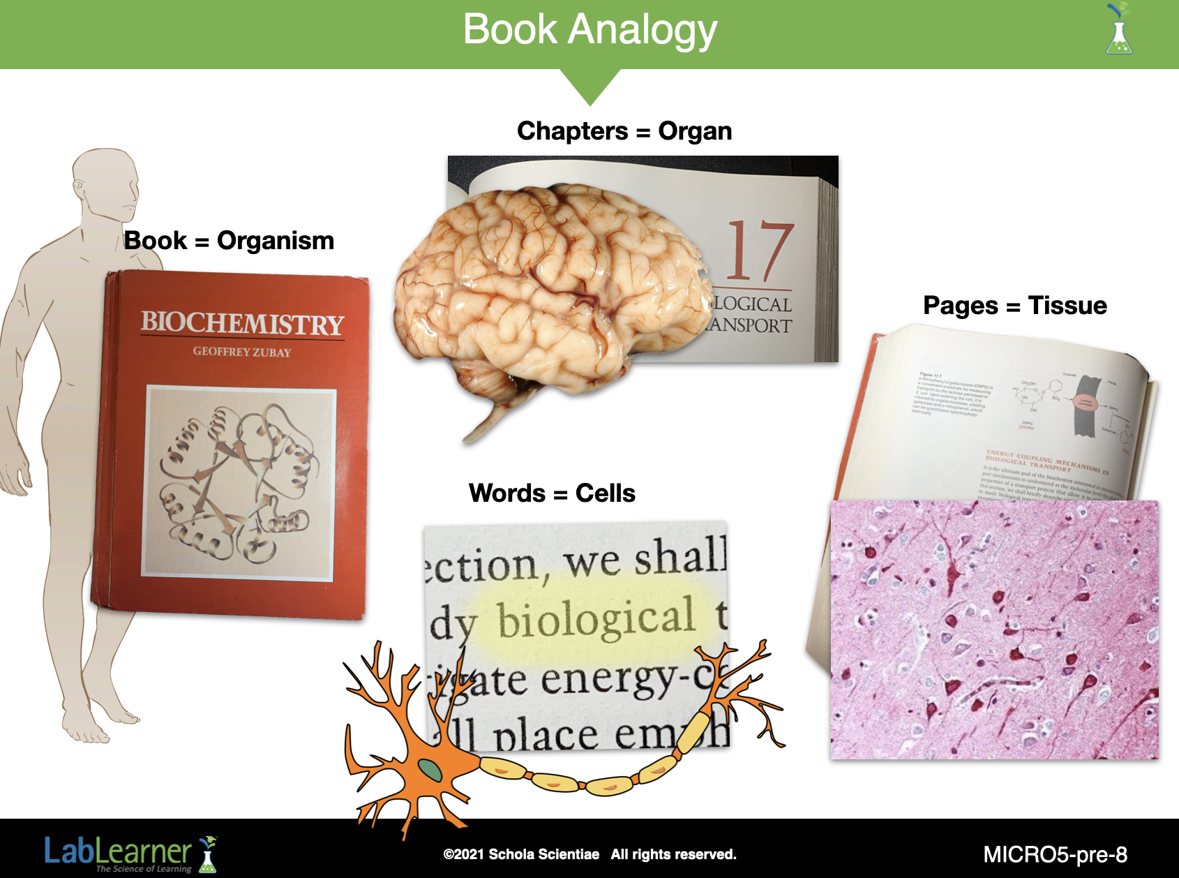
Use an analogy to facilitate student comprehension of the relationship between cells, tissues, organs, and organisms. Divide students into groups of four. Explain that you would like them to brainstorm and think of an analogy that describes the relationship between cells, tissues, organs, and organisms. If necessary, discuss with students what is meant by the term analogy.
To help students, share an example of an analogy:
Organism = Book
Organs = Chapters
Tissues = Pages
Cells = Words
Students should record their examples in Problem 1 of their Scientist Data Record.
______________________________________________
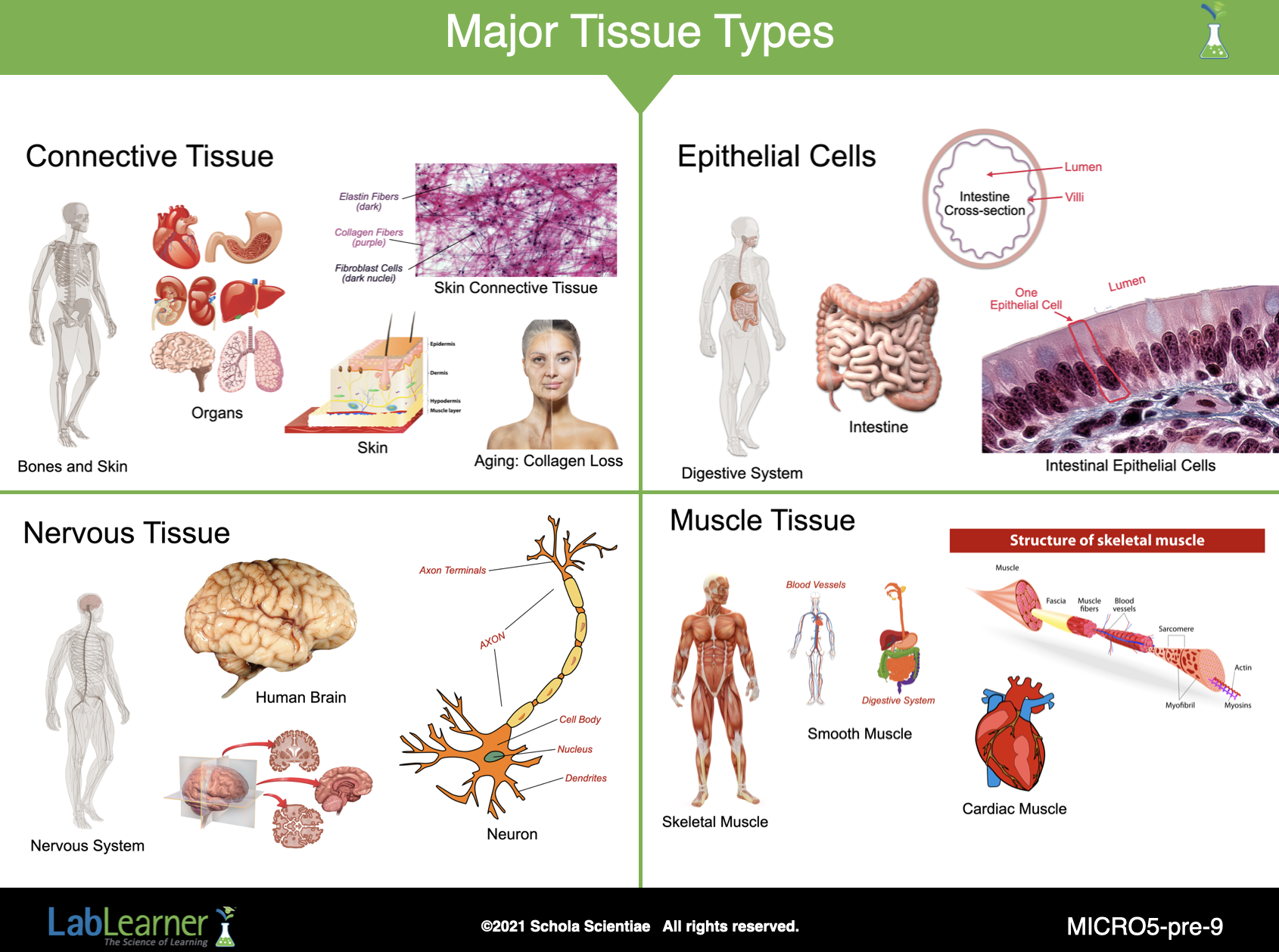
SLIDE MICRO5-pre-9
This slide simply shows the four different major types of tissue in a human (or another animal) body.
- Connective Tissue
- Epithelial Tissue
- Nervous Tissue
- Muscle Tissue
Each section of the slide illustrates the various levels of organization of tissue and organs, as well as organ systems.
Next, tell students that in Investigation Five they will observe slides of four different specimens. The slides are of different tissues, structures, and organs of plants and animals.
Explain that several of the specimens they will view differ from those that they observed in Investigations Two through Four in the way in which they were prepared.
Ask students: Did you prepare the fingerprint, cheek cell, and onion specimens in Investigations Two, Three, and Four the same way? Students should indicate that different procedures were used for specimen preparation in Investigations Two, Three, and Four.
Ask students: Can you name different ways in which you prepared a specimen for analysis? Students should indicate that a paper with a fingerprint was placed onto the slide in Investigation Two. In Investigations Three and Four, a sample of cheek cells and onion skin was placed in water on a slide.
Help students recall and classify three different ways in which they prepared a specimen: whole-mount, smear (type of wet mount), and section (type of wet mount). The fingerprint specimen in Investigation Two was prepared as a whole mount: the entire piece of paper with the fingerprint was placed onto the slide. The cheek cell specimen was prepared as a smear, a type of wet mount: a sample was taken from the cheek and smeared onto the water on the slide. The onion specimen was prepared as another type of wet mount: a slice or section. A piece of onion skin was removed from the whole onion and placed onto the water on the slide.
Ask students: Why was it necessary to prepare a piece of the onion for analysis and smear of the cheek cells? Why could the “whole mount” procedure not be used for these two specimens? Students should suggest that a whole-mount procedure involves placing the entire structure on a slide. This would not be possible with the cheek or a whole onion. Neither would fit on a slide or under the objectives of the microscope.
______________________________________________
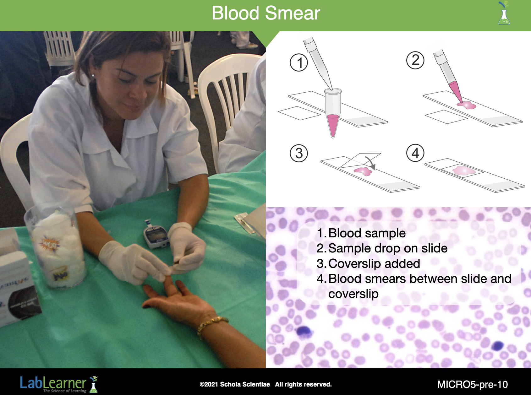
SLIDE MICRO5-pre-10
With this slide, we begin to prepare for Investigation 5 lab. In the lab, students will examine four prepared slides that were prepared by three different procedures:
- Smear: (human blood)
- Whole Mount: (Elodea leaf) Similar to a wet mount preparation that students prepared in Investigation 4.
- Cross-Section: (Pinus [pine] stem) and human colon (large intestine)
SLIDE MICRO5-pre-10 illustrates how blood samples are prepared for microscopic examination. The micrograph (photograph taken through a microscope) in the background shows nicely smeared red blood cells (erythrocytes) as well as two darkly stained white blood cells (leukocytes).
NOTE THAT THE PRELAB EXERCISE INVOLVING MODELING OF CROSS-SECTIONS AND LONGITUDINAL SECTIONS IS OMITTED FROM THIS PRELAB. THIS INVOLVES STUDENT SDR PAGES 76 (BEGINNING WITH PROBLEM 2) THROUGH 80.
______________________________________________
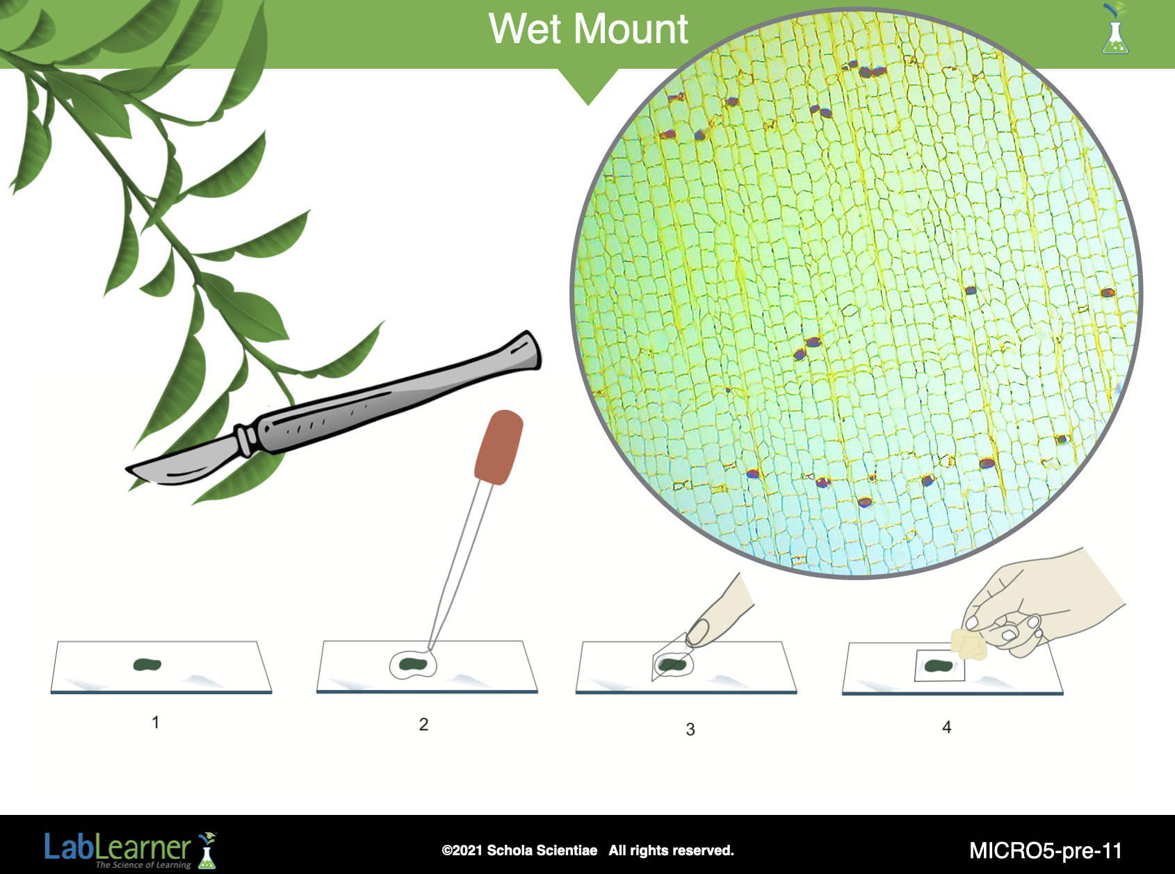
SLIDE MICRO5-pre-11
This slide shows the process used to prepare a wet mount. The whole mount of an Elodea leaf that students will examine in Investigation Five lab is prepared in a similar manner except that the specimen is dry and permanently fixed between the slide and coverslip.
______________________________________________
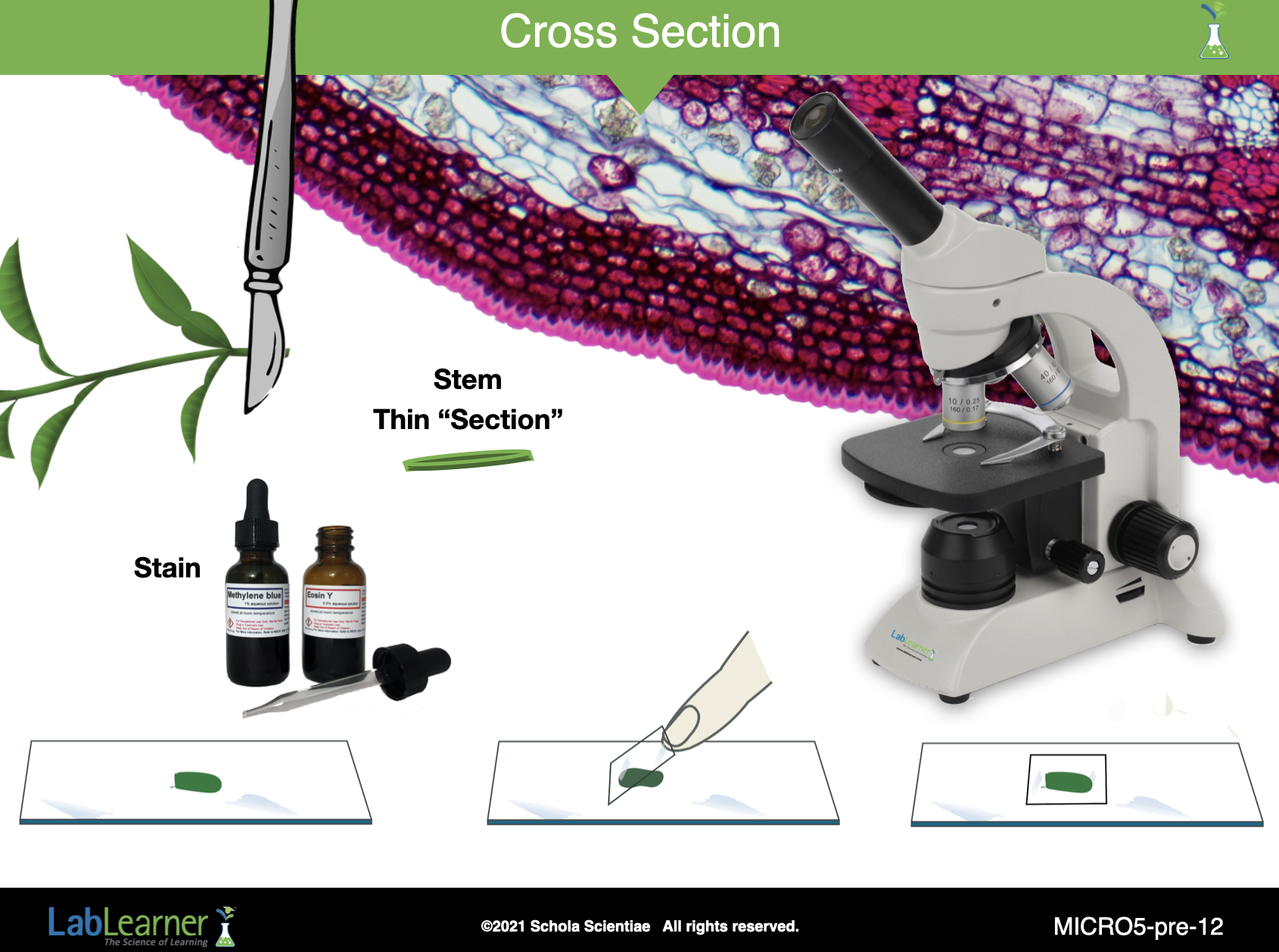
SLIDE MICRO5-pre-12
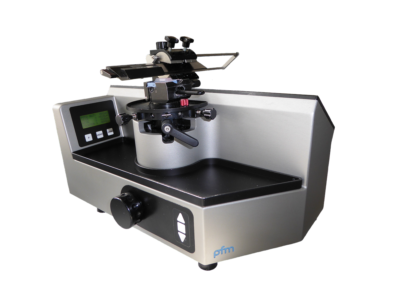
This slide shows how cross-section slides are prepared. while it shows the cross-cutting (sectioning) of a plant stem, in reality, most cross-sections are prepared with the use of an instrument called a microtome, that can cut extremely (one cell think) sections. A microtome is shown below:
______________________________________________
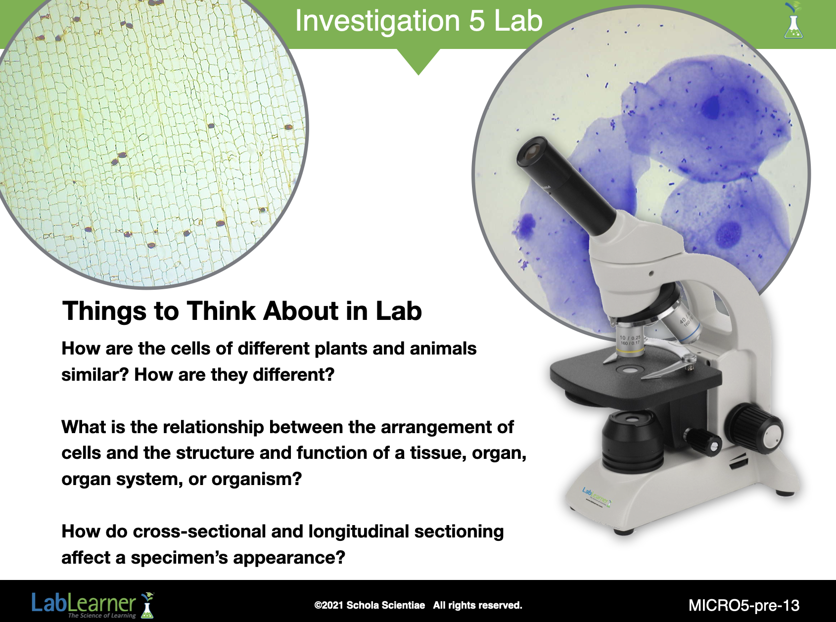
SLIDE MICRO5-pre-13
This final slide simply asks students to consider three questions as they examine their prepared sides in the Lab:
How are the cells of different plants and animals similar? How are they different?
What is the relationship between the arrangement of cells and the structure and function of a tissue, organ, organ system, or organism?
How do you think cross-sectional and longitudinal sectioning might affect a specimen’s appearance?
______________________________________________
WATCH IT
Play the following Student Video in preparation for the lab. Discuss as necessary to answer student questions.