Teacher Portal:
Microscopic Explorations
Investigation 5 – PostLab
ASK WHY
Microscopes are one of the most important scientific instruments developed. In fact, in the medical field, microscopes are largely responsible for making modern medicine “modern”!
BRANCH OUT
You might wonder which came first, the microscope or the telescope. Actually, they appear to have appeared at about the same time although it is thought that the microscope may have come first. In either case, the first forms of both instruments were developed in the 16th century (the n1500s). It isn’t that much of a stretch to imagine that, once the microscope was invented, that someone would consider making similar instruments that could make distant objects appear closer.
PRINT IT
Use your browser to download a printable PDF as help during the slide presentation and to make additional notes. In your browser, go to File > Print and then choose to save as PDF.
NAVIGATE IT
Once the slide presentation is launched
- use your left and right arrows to advance or go back in the slide presentation, and
- hover your mouse over the left edge of the presentation to get a view of the thumbnails for all the slides so that you can quickly move anywhere in the presentation.
- Click HERE to launch the slide presentation for the CELL.
SHARE IT
SLIDE MICRO5-post-1
Begin this part of the investigation by reviewing the experiments students performed in the lab. Ask the following questions to promote discussion of the experiments.
What was the theme or focus of the investigation? The theme or focus of the investigation was to investigate how cells are arranged in different plant and animal tissues and organs. In addition, students examined how the preparation (sectioning, staining) of specimens can affect what is seen when viewing the specimen under the microscope.
Think about Trial 1. What questions did you answer when performing Trial 1? How did you answer these questions? Students investigated the questions: How are the cells of different plants and animals similar? How are they different? What is the relationship between the arrangement of cells and the structure and function of a tissue, organ, or organism? In order to answer the question, students viewed an Elodea leaf, a Pinus Stem, a Blood smear, and a Colon specimen with the compound microscope.
What question was Trial 2 designed to answer? How did you answer this question? Trial 2 was designed to answer: How do cross-sectional and longitudinal sectioning affect a specimen’s appearance? Students answered the question by building a model of the colon from gram cubes and simulating the preparation of a cross-section and longitudinal section of the colon model.
______________________________________________
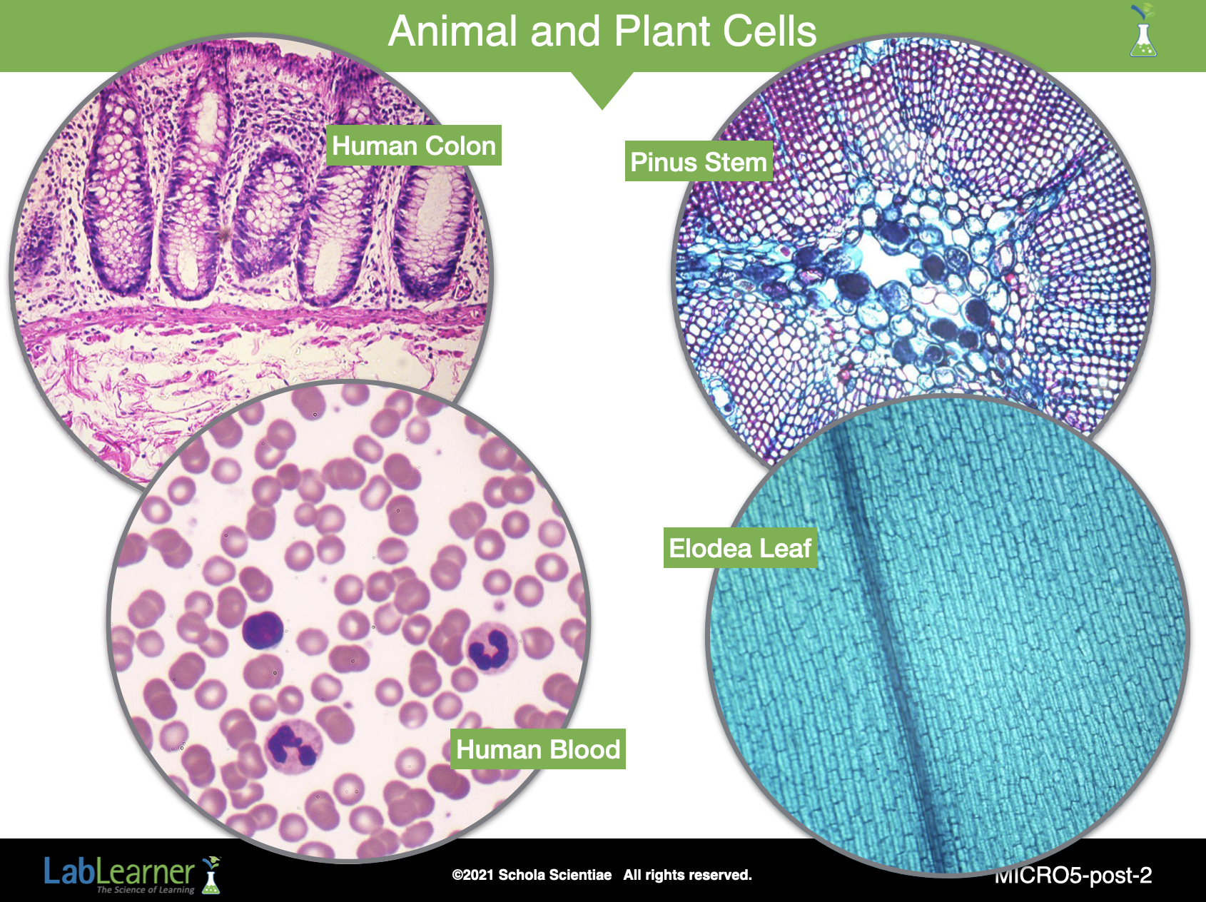
SLIDE MICRO5-post-2
Begin analysis of Trial 1 by discussing students’ observations of the four specimens: the Elodea leaf, the Pinus Stem, the human blood smear, and the human colon.
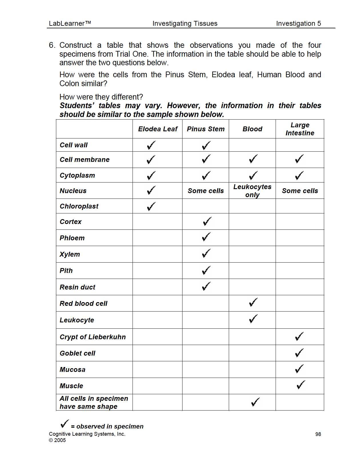
Ask students: You viewed four different specimens during the Lab. Two were from plants and two were from animals. How would you construct a table using the observations from Trial One to help answer the questions: How were the cells in each specimen similar? How were they different? Students’ answers should reflect an understanding of the Procedural Tool, Creation and Completion of a Data Table, that they used in earlier investigations. If students require assistance, ask the following questions as prompts. Encourage students to work with a partner to create a table that compiles the observations made during their investigation. If students have difficulty with the task, work on the process as a class. Students should record their table in problem 6 of their Scientist Data Record.
Use the following questions to guide students as they complete the table.
- Did you observe cells in both specimens with a cytoplasm, cell wall, and cell membrane?
- Were the cells in the Elodea specimen all the same shape?
- Were the cells in the Pinus Stem specimen all the same shape?
- Did the cells of the Elodea leaf and Pinus Stem have the same shape?
- Did you observe cells in the Blood smear and Colon with cytoplasm and cell membrane?
- Were the cells in the blood specimen all the same shape?
- Were the cells in the colon specimen all the same shape?
- Did the cells of the blood and colon specimen have the same shape?

______________________________________________
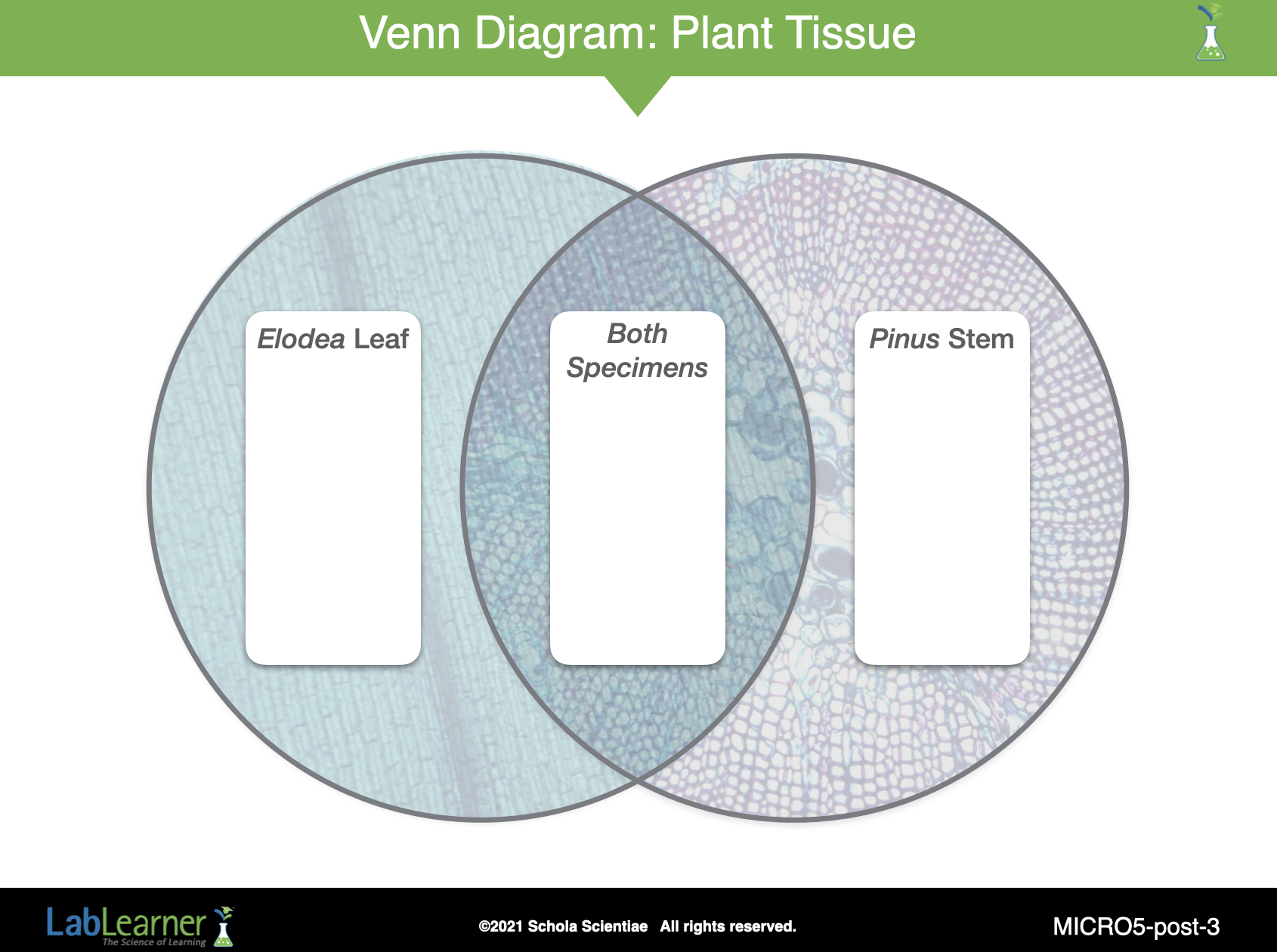
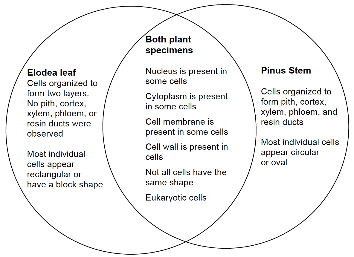
SLIDE MICRO5-post-3
Even after answering the smaller questions, it may be difficult for students to summarize their answers from the wealth of data in the table. To help students with their summary suggest that students use a Venn diagram to organize their results.
a. Briefly review the structure of a Venn diagram and how it can be used to contrast the differences and similarities of things that are to be compared.
b. Encourage the class to help you complete a Venn diagram for the plant specimens using the data from their data table.
______________________________________________
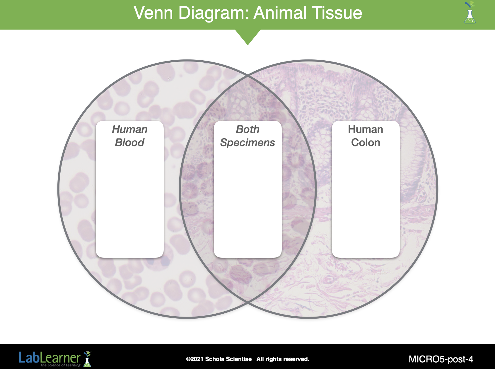
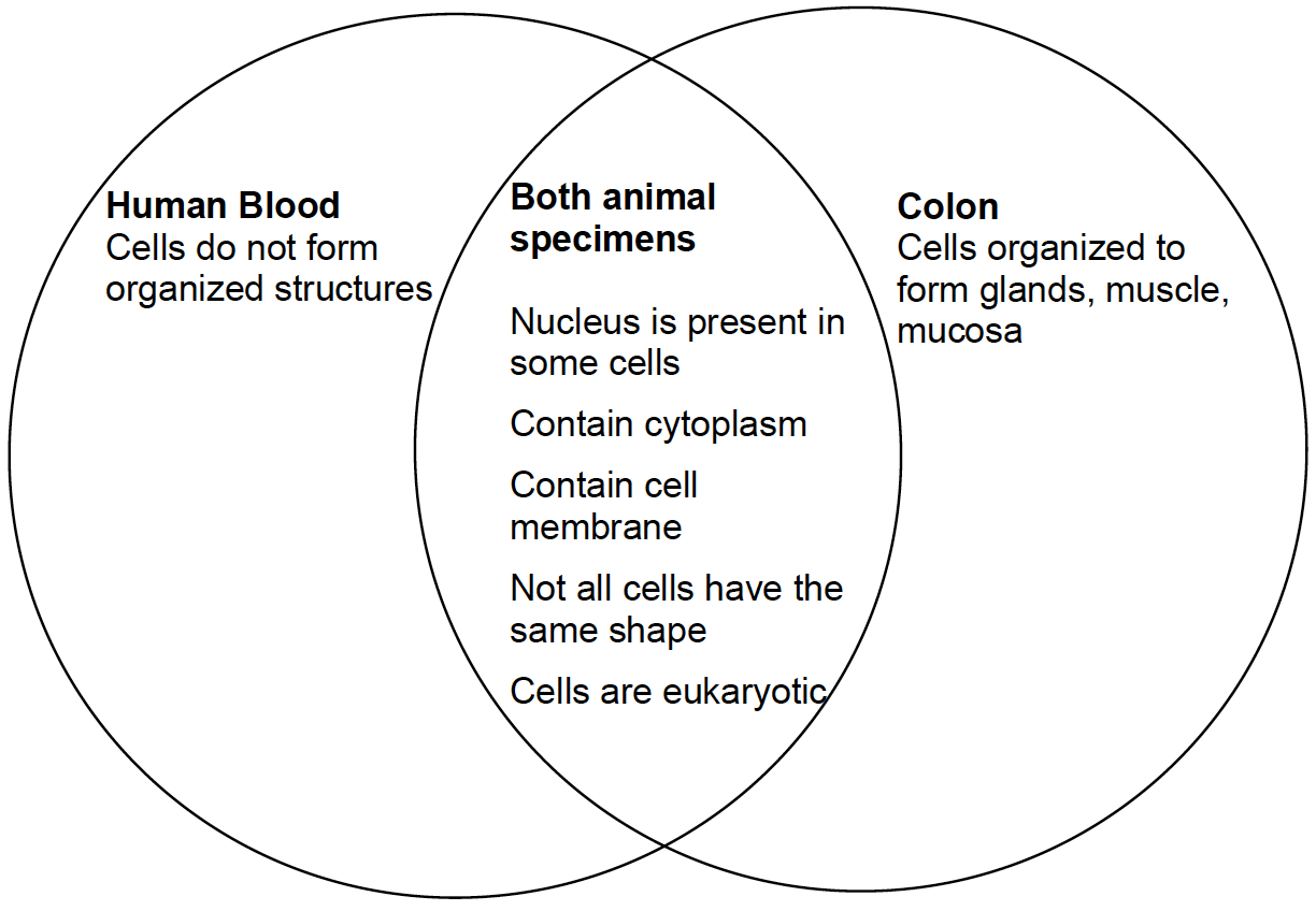
SLIDE MICRO5-post-4
Encourage the class to help you complete a Venn diagram for the animal specimens using the data from their data table.
______________________________________________
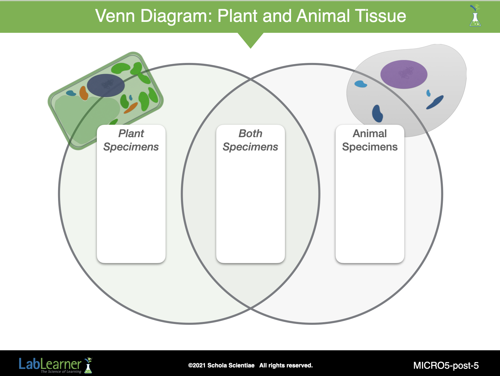
SLIDE MICRO5-post-5
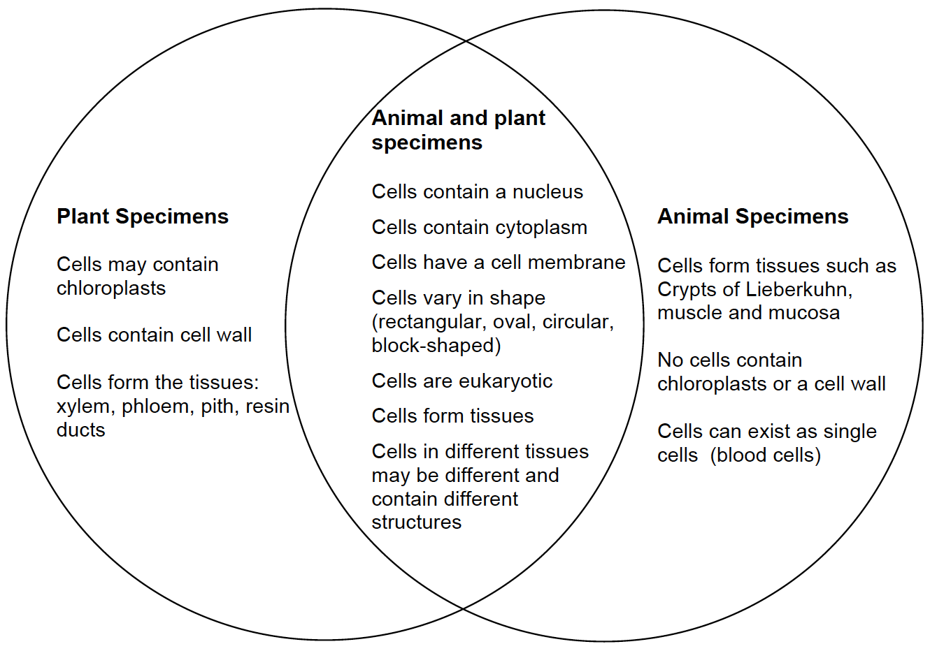
Ask students: How could you combine the information from both Venn diagrams to make a Venn diagram comparing plant and animal specimens? Student answers may vary. Help students combine the information from the Plant Venn diagram and Animal Venn diagram to create a new one that compares cells from plant and animal specimens.
Ask students: Think of the cheek cells and onion cells you observed in Investigations Three and Four. Does the Venn diagram accurately reflect your observations from the cheek and onion cells? Students should indicate that the Venn Diagram does reflect their observations of the cheek and onion cells. Observations of the onion and cheek cells support the finding that animal and plant cells can vary in shape, both contain a nucleus, cytoplasm, and cell membrane.
After the Venn diagram is complete, ask for one or two student volunteers to summarize the differences and similarities between the plant and animal specimens in several sentences. Encourage students to record their summaries in Problem 7 of their Scientist Data Record.
______________________________________________
SLIDE MICRO5-post-6
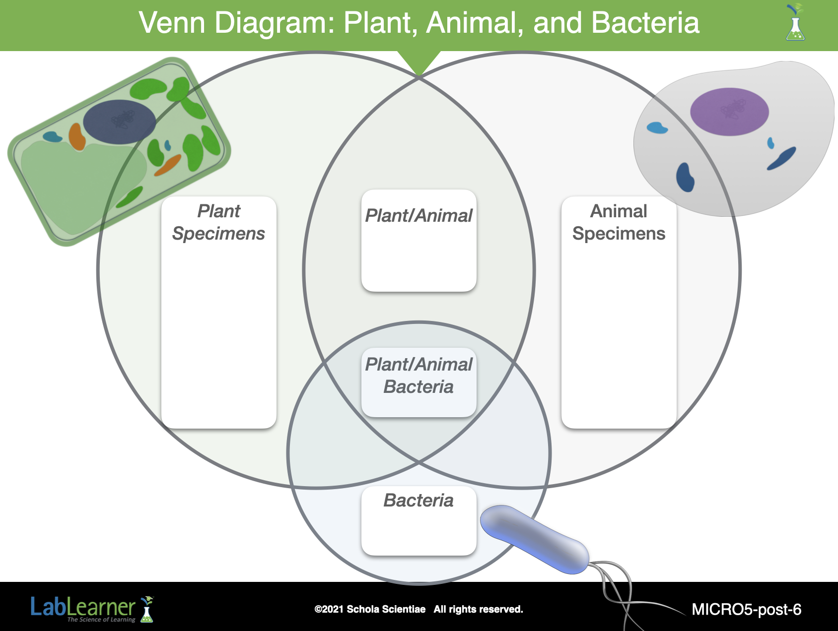
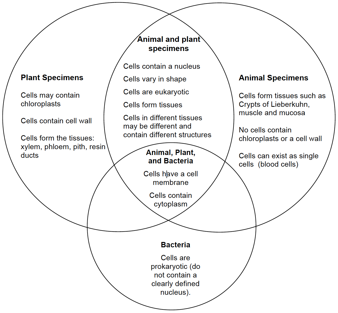
Ask students: In Investigation Three you learned about three types of cells: animal cells, plant cells, and bacterial cells. You observed specimens of plant and animal cells and read a passage about bacterial cells. How could you change your Venn Diagram to include information about bacterial cells? Student answers may vary as students have not had much experience viewing bacterial cells. However, using the information from the reading passage in Investigation Three and the definition of prokaryotic cells in their Scientist’s Glossary students should be able to construct a Venn Diagram using the small amount of information they have gained from these sources.
______________________________________________
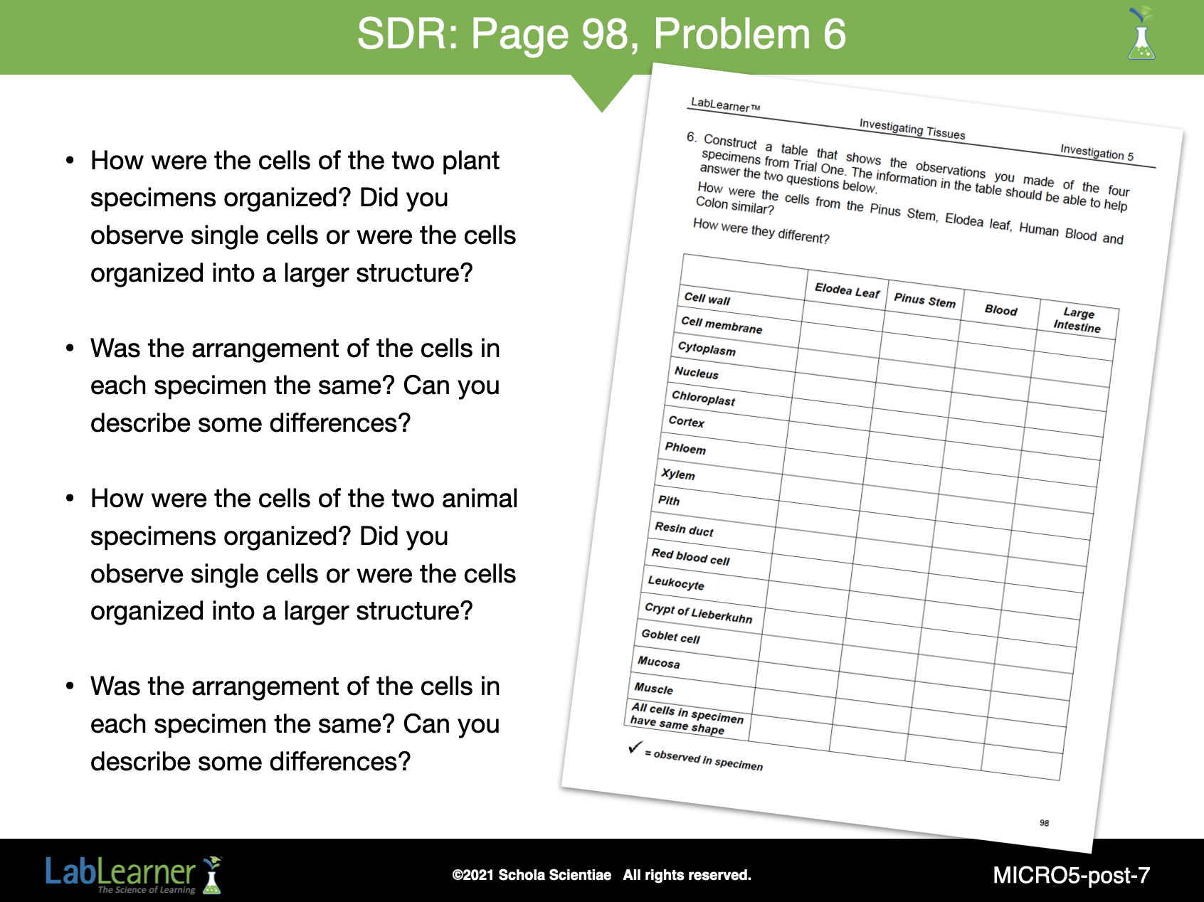
SLIDE MICRO5-post-7
Continue the analysis, focusing on the relationship between structure and function that students were to investigate during Trial One.
Ask students: The second question you investigated in Trial One was: What is the relationship between the arrangement of cells and the structure and function of a tissue, organ, or organism? What information could you add to your table to help you answer this question? Students should suggest adding two items to their tables:
- Whether single cells were present in the specimen
- Whether cells were organized in layers or tissues
If necessary, use the following questions to assist students in developing their answers. Direct students to add the additional information to the table they completed in Problem 6 of their Scientist Data Record.
- How were the cells of the two plant specimens organized? Did you observe single cells or were the cells organized into a larger structure?
- Was the arrangement of the cells in each specimen the same? Can you describe some differences?
- How were the cells of the two animal specimens organized? Did you observe single cells or were the cells organized into a larger structure?
- Was the arrangement of the cells in each specimen the same? Can you describe some differences?
______________________________________________
KEYS: