Teacher Portal:
Microscopic Explorations
Investigation 4 – PostLab
ASK WHY
Microscopes are one of the most important scientific instruments developed. In fact, in the medical field, microscopes are largely responsible for making modern medicine “modern”!
BRANCH OUT
You might wonder which came first, the microscope or the telescope. Actually, they appear to have appeared at about the same time although it is thought that the microscope may have come first. In either case, the first forms of both instruments were developed in the 16th century (the n1500s). It isn’t that much of a stretch to imagine that, once the microscope was invented, that someone would consider making similar instruments that could make distant objects appear closer.
PRINT IT
Use your browser to download a printable PDF as help during the slide presentation and to make additional notes. In your browser, go to File > Print and then choose to save as PDF.
NAVIGATE IT
Once the slide presentation is launched
- use your left and right arrows to advance or go back in the slide presentation, and
- hover your mouse over the left edge of the presentation to get a view of the thumbnails for all the slides so that you can quickly move anywhere in the presentation.
- Click HERE to launch the slide presentation for the CELL.
SHARE IT
SLIDE MICRO4-post-1
Begin this part of the investigation by reviewing the experiments students performed in the lab. Ask the following questions to promote discussion of the experiments.
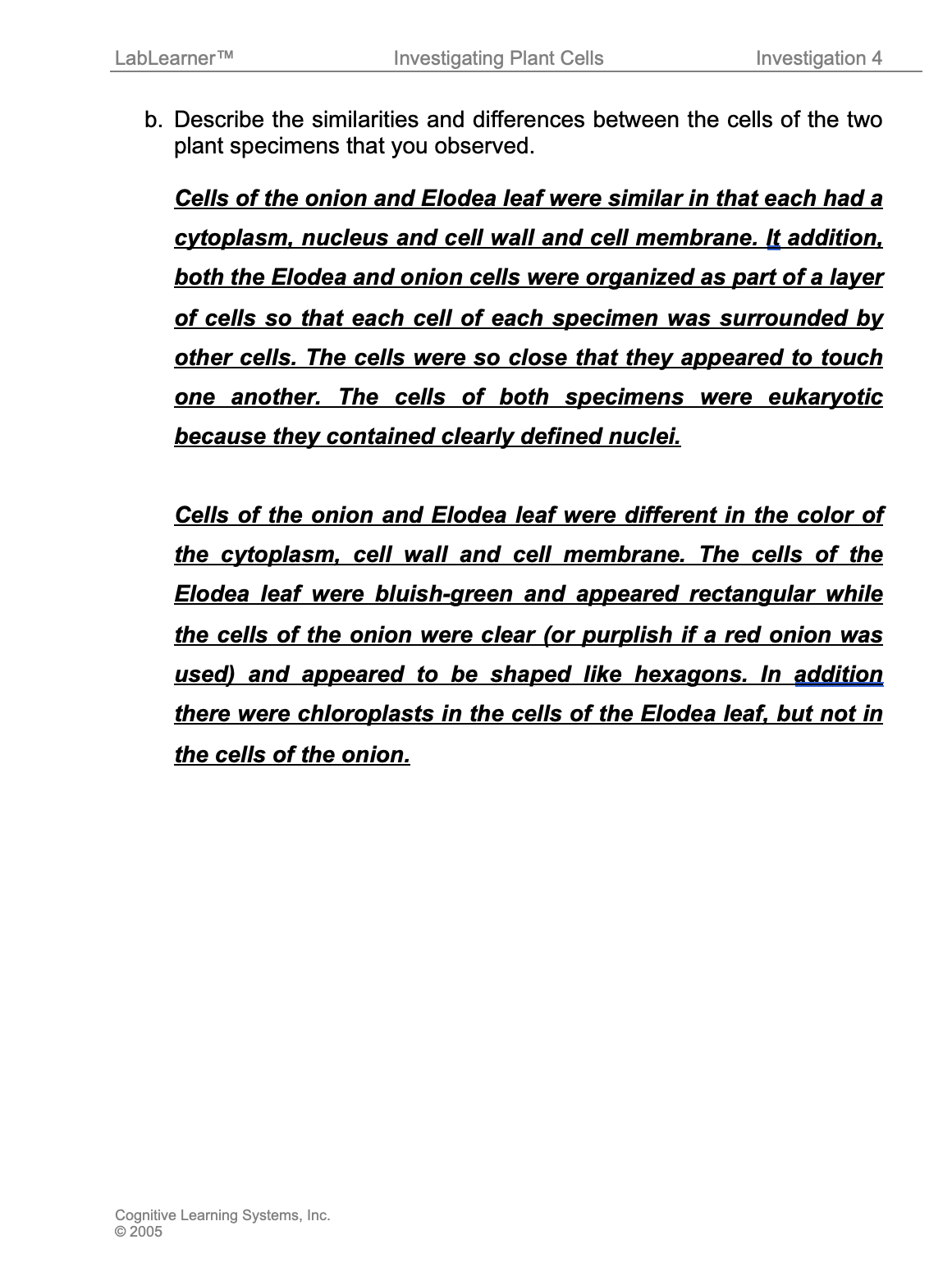
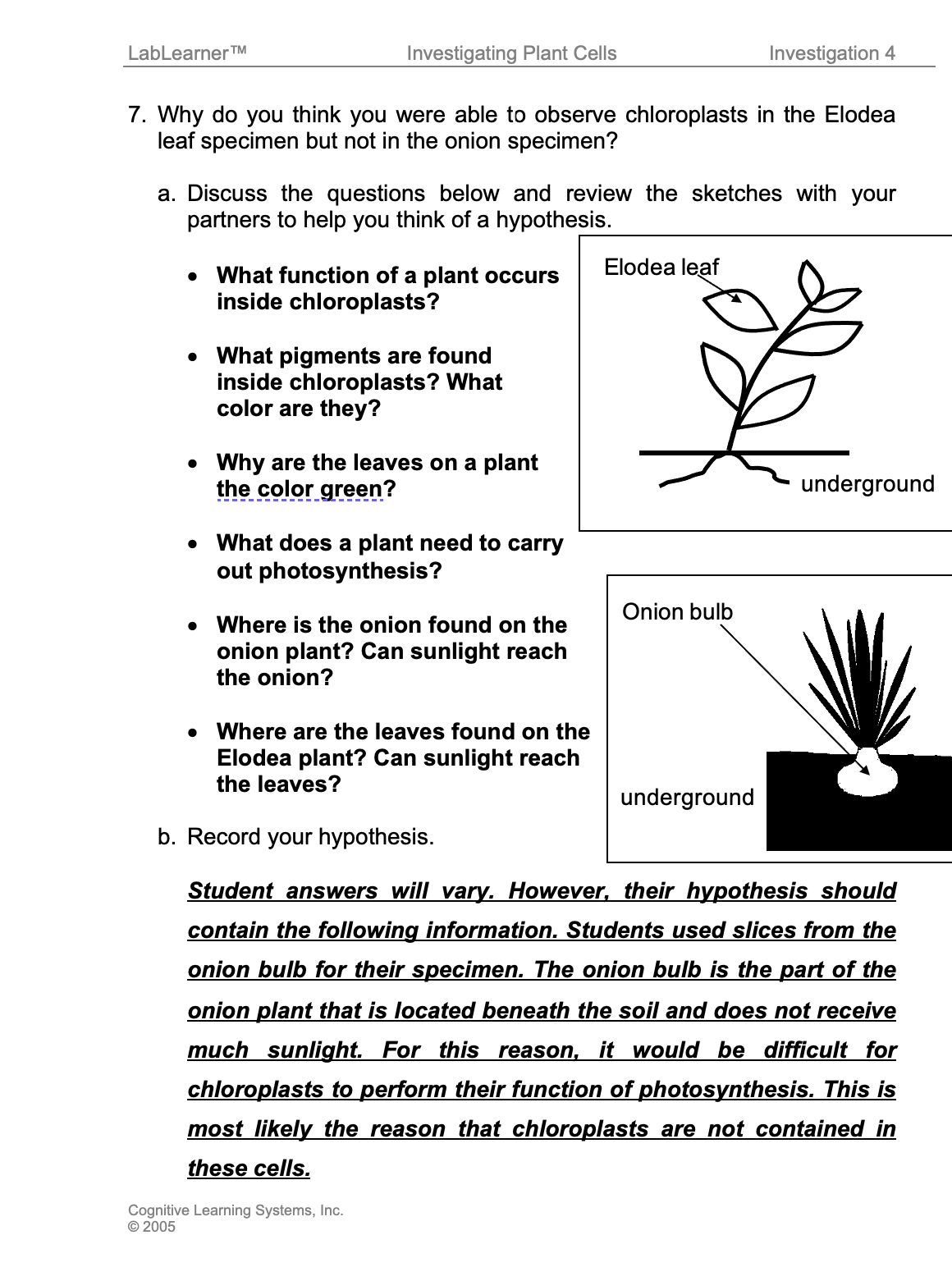
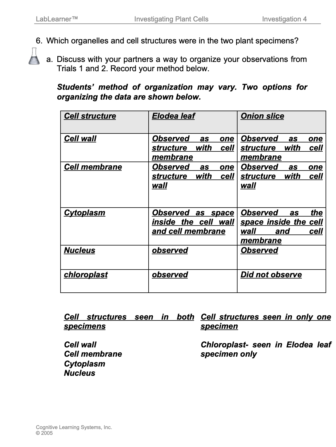
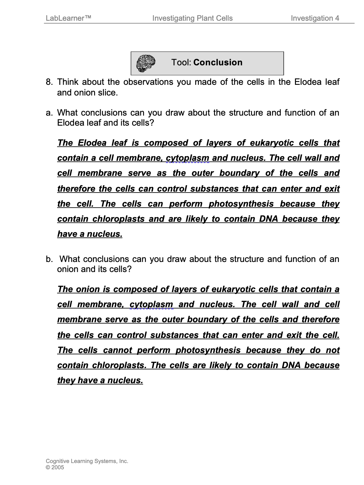
What was the theme or focus of the investigation? The theme or focus of the investigation was to investigate the structures found in plant cells. Students investigated the appearance of plant cells by observing a prepared slide of an Elodea leaf and by preparing a wet mount of an onion slice.
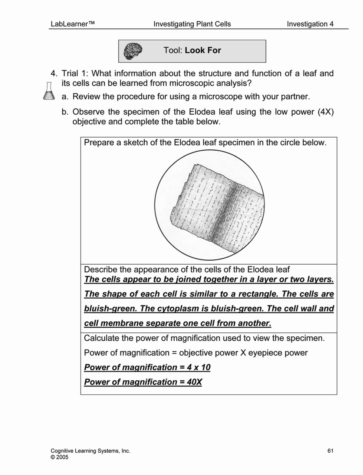
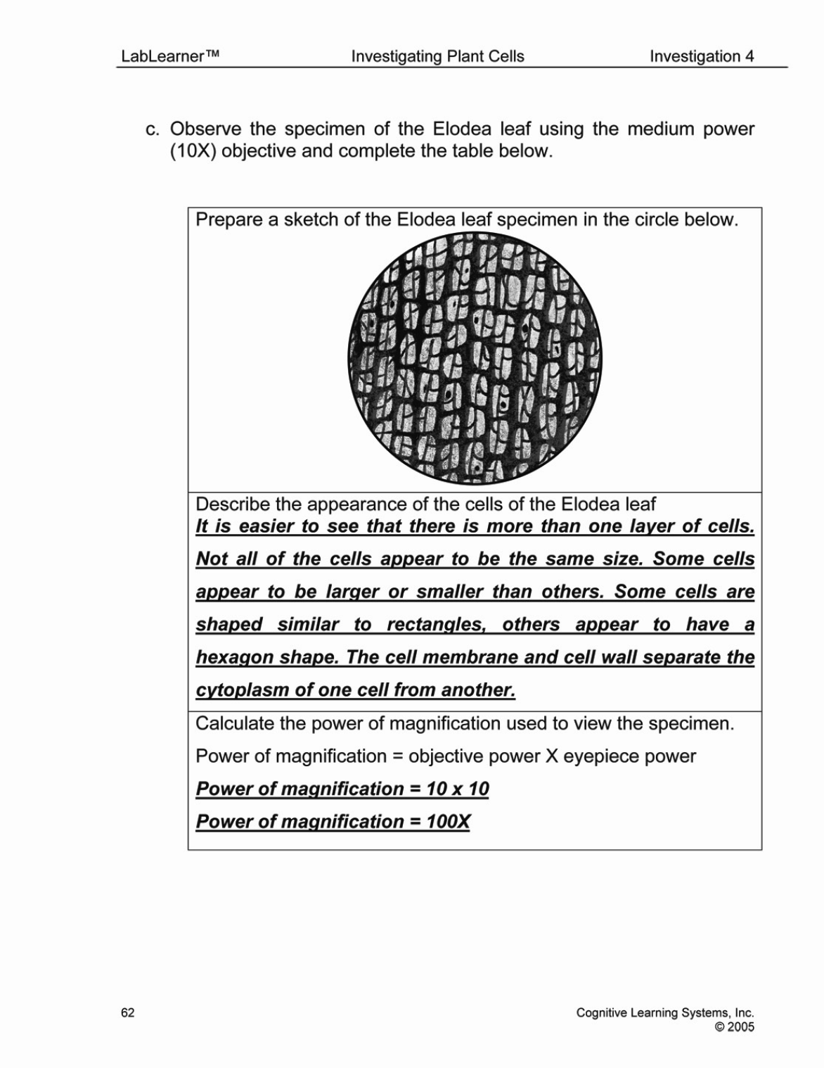
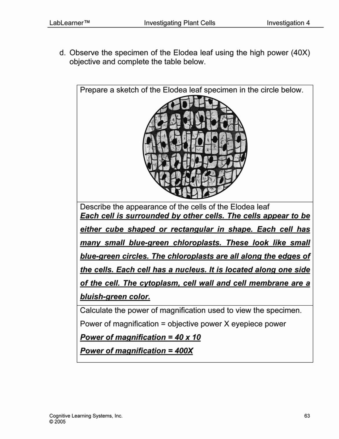
In Trial 1, you observed an Elodea leaf using the microscope. What question did you answer when performing Trial 1? How did you answer this question? Students investigated the question: What information about the structure and function of a leaf and its cells can be learned from microscopic analysis? To answer this question, students viewed a prepared slide of an Elodea leaf using the compound microscope. Students identified the cell organelles and used drawings and written descriptions to record their observations.
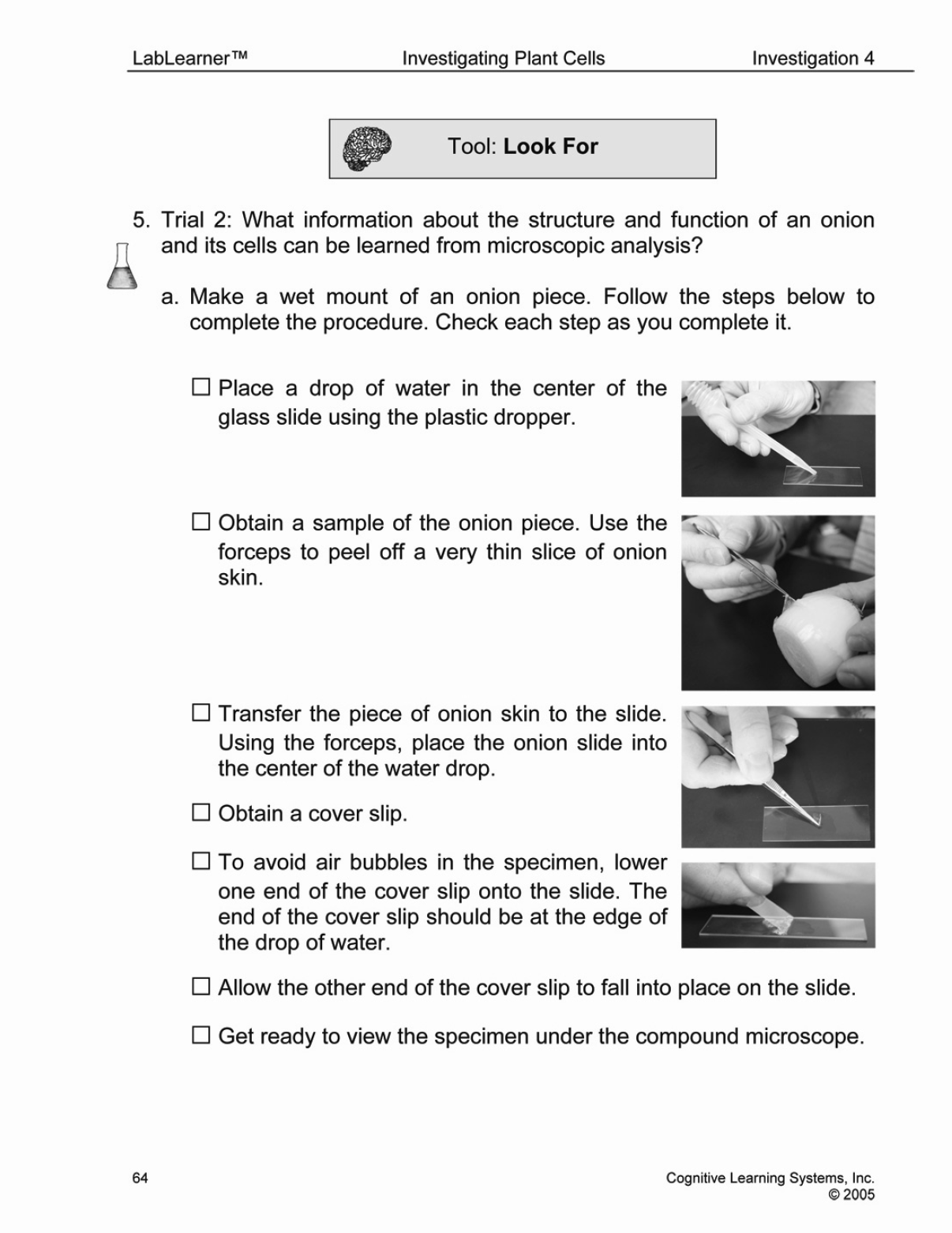
In Trial 2, you created and viewed a wet mount slide of an onion. What question was Trial 2 designed to answer? How did you answer this question? Trial 2 was designed to answer: What information about the structure and function of an onion and its cells can be learned from microscopic analysis? To answer this question, students prepared a wet mount of an onion slice and viewed the onion slice with the compound microscope. Students identified the cell organelles and used drawings and written descriptions to record their observations.
______________________________________________
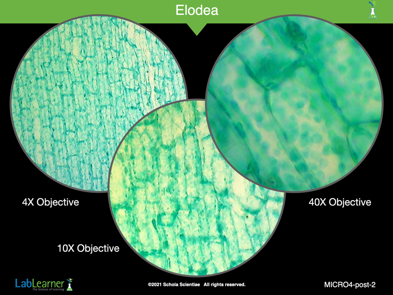
SLIDE MICRO4-post-2
Begin analysis of Trial 1 by discussing students’ observations of the Elodea leaf.
Ask students: In Trial One you viewed the Elodea leaf under three different objectives. Can you describe some of the differences and similarities you observed using the three different objectives? Student answers may vary. However, student answers should discuss the following similarities: the color and proportions of the Elodea leaf cells were similar when viewed with the two objectives. In other words, the different lenses did not produce a change in color or a change in the proportions of the images. Students should have observed that the cells appeared to be in direct contact with other cells regardless of the objective used.
As the level of magnification increased, students will have been more likely to observe the nucleus and chloroplasts in each cell. Students should indicate that it was difficult to locate the chloroplasts and nucleus using the 4X and 10X objectives, but that they could be seen when using the 40X objective. As the magnification increased, it should also have been easier for students to discern the exact shape of each cell. However, the overall structure of the leaf and the arrangement of cells in a layer should have been easier for students to observe when using the 4X objective.
______________________________________________
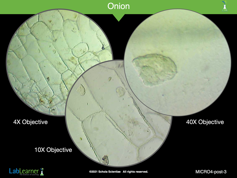
SLIDE MICRO4-post-3
Continue the analysis of Trial 2 by discussing students’ observations of the onion slice.
Ask students: In Trial Two you viewed an onion slice under three different objectives. Can you describe some of the differences and similarities you observed using the three different objectives? Student answers may vary. However, student answers should discuss the following similarities: the color and proportions of the onion cells were similar when viewed with the two objectives. In other words, the different lenses did not produce a change in color or a change in the proportions of the images. Students should have observed that the cells appeared to be in direct contact with other cells regardless of the objective used.
As the level of magnification increased, students will have been more likely to observe the nucleus in each cell. Students should indicate that it was difficult to locate the nucleus using the 4X objective, but that these could be seen when using the 10X and 40X objectives. As the magnification increased, it should also have been easier for students to discern the exact shape of each cell. However, the overall structure of the onion skin and the arrangement of cells in a layer should have been easier for students to observe when using the 4X objective.
______________________________________________
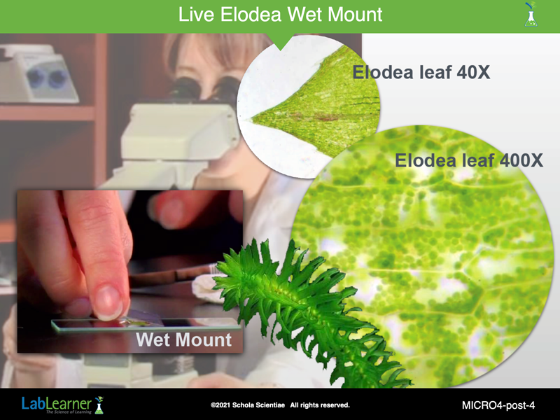
SLIDE MICRO4-post-4
This slide is from the LabLearner middle school CELL Photosynthesis. It is included here to show students that sometimes living tissue can give a better picture upon microscopic examination.
Notice how bright green the chloroplasts are in the 400X image. A sprig of living Elodea is shown at the lower center.
Students may be interested to note that techniques such as the preparation of a wet mount slide will be used again and again in the future as they progress through the LabLearner curriculum. The teacher may point out the binocular, oil immersion microscope show in the background of this slide. Students will use this type of microscope in middle school LabLearner.
______________________________________________
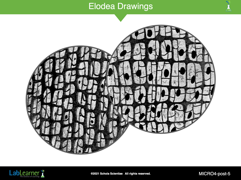
SLIDE MICRO4-post-5
Ask students: Did your sketches accurately represent the images seen with each objective? Were the sketches drawn as close to scale as possible? Did the sketches represent the change in the power of magnification seen with each objective? Student answers should reflect the understanding that accurate sketches attempt to maintain the proportions of the image viewed with the microscope. In addition, the sketches made with different powers of magnification should portray those differences as accurately as possible. For example, the structures in the sketches of the 40X objectives should be larger than those in the sketches of the specimen from the 10X objective.
- Encourage several student volunteers to illustrate the points discussed above by drawing their series of sketches on the board.
- Discuss how the sketches maintain the proportions of the images and how the sketches attempt to accurately represent the changes in scale seen with each objective.
- If students’ sketches were not accurate representations, encourage them to predict how each sketch would appear if the points described above were taken into consideration. Draw the new sketches on the board.
Ask students: How do you think the images of the cells that you drew compare to the actual size of a plant cell? Why? Students should indicate that an actual plant cell is smaller than the images seen with the compound microscope because the microscope produced magnified images of specimens.
______________________________________________
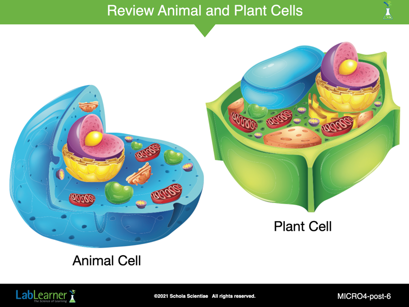
SLIDE MICRO4-post-6
This slide simply shows both an animal cell and a plant cell next to each other for comparison. Look at all of the similarities and differences.
______________________________________________
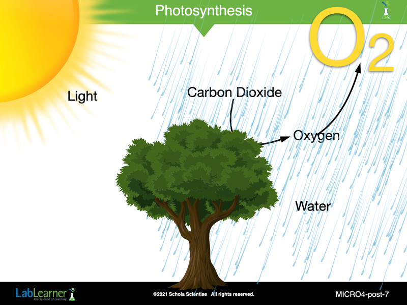
SLIDE MICRO4-post-7
This slide brings us back to the photosynthesis reaction and accentuates the importance of oxygen production by plants.
______________________________________________
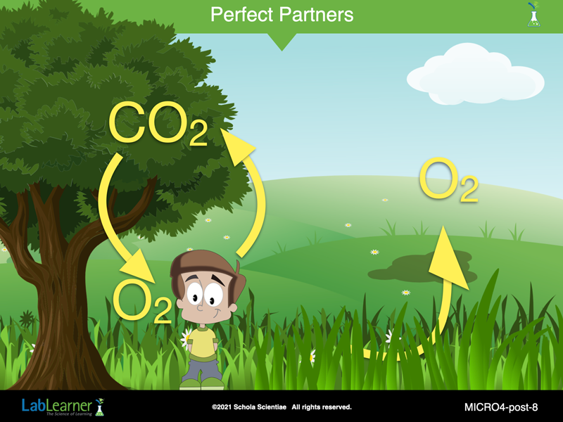
SLIDE MICRO4-post-8
This slide shows the dependence that we, as humans, have on plants. When we breathe out we exhale carbon dioxide (CO2). This gas is lethal to animals, we need oxygen to survive. Fortunately, plants need CO2 as a part of the photosynthesis reaction along with water. They convert CO2, in the presents of light and water, into oxygen (O2) for us to breathe. What a wonderful partnership!
______________________________________________

SLIDE MICRO4-post-9
This final slide simply stresses the massive amount of oxygen that is produced by plants on Earth. If it was not for the photosynthesis carried out by plants, there would be no animal life (including humans) on this planet.

______________________________________________
PRELAB EXERCISE
Divide students into the five cooperative groups from the Lab. Encourage student groups to develop a way to organize their data. Students should record their method in problem 6a of their SDRs. The following are guiding questions that students should ask themselves as they create their method of organizing the data.
-
-
- What type of results or information did I obtain?
- How could I combine the data I recorded for each trial I conducted in the Lab?
- Are there common features of the specimens?
-
After students have had an opportunity to work with organizing their data, ask for one of the groups to share their method of organization with the class by reconstructing it on the board. Student choices for data organization may vary but should contain the following information.
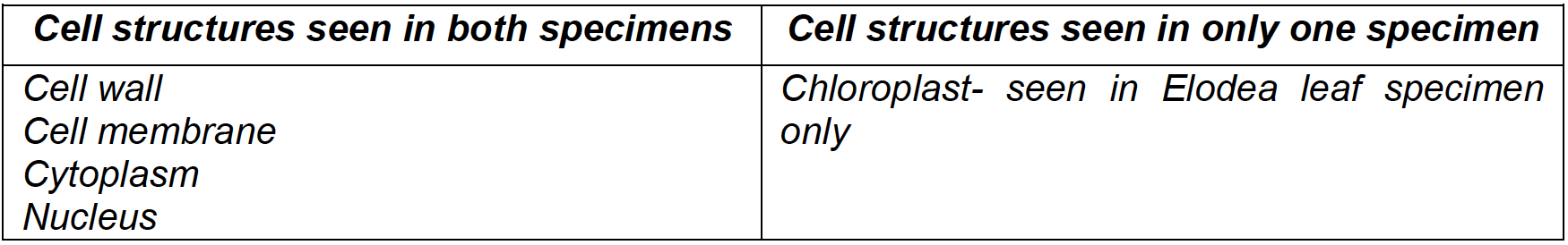
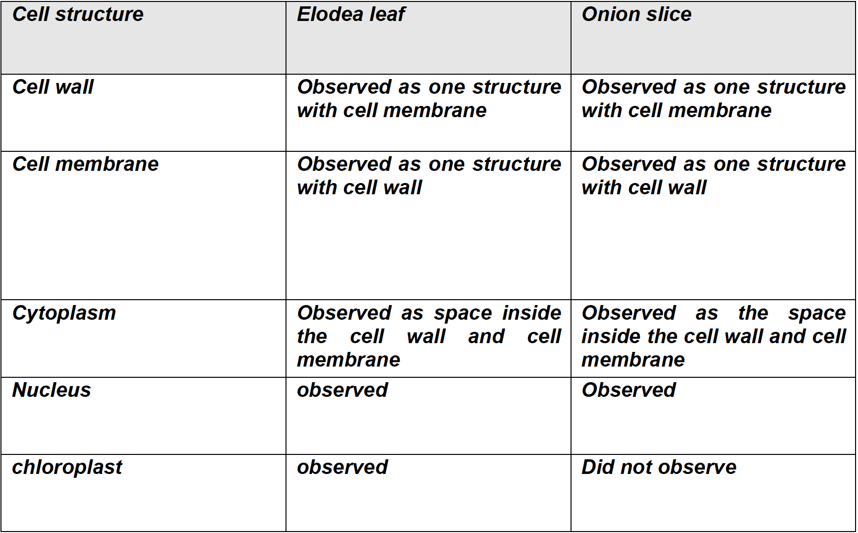
Encourage students to use the information they organized to answer the question: Did you observe the same organelles and cellular structures in the cells from both plants? Students should indicate that the cell wall, cell membrane, cytoplasm, and nucleus could be seen in both the Elodea leaf and onion slice specimens. Chloroplasts could only be seen in the Elodea leaf specimen. However, some students may indicate that they did not see nuclei or the cell wall and cell membrane in both specimens. Use the following questions and the Elodea and Onion Specimen Transparency to help students clarify their observations.
KEYS: