Teacher Portal:
Microscopic Explorations
Investigation 4 – Lab
ASK WHY
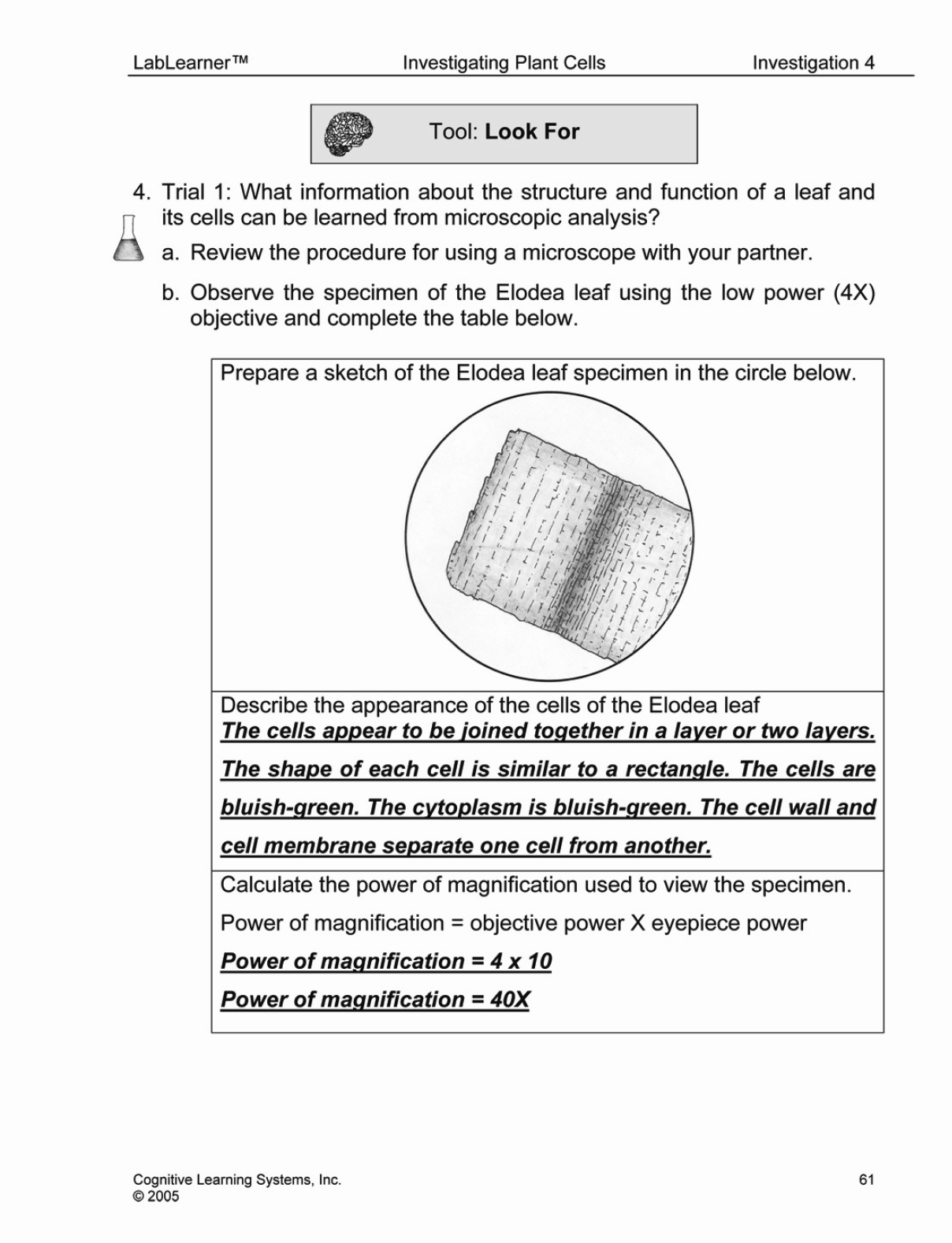
Microscopes are one of the most important scientific instruments developed. In fact, in the medical field, microscopes are largely responsible for making modern medicine “modern”!
BRANCH OUT
In LabLearner we use compound light microscopes. These are very widely used in both research and clinical and industrial applications. However, there are many additional types of microscopes that are used for various purposes. Among the most useful in research is the electron microscope. While light microscopes have a maximum magnification of about 1,000X, electron microscopes have magnifications of around 300,000X with even better resolution than a light microscope. Whereas light microscopes may be able to see organelles within cells, like the nucleus and even mitochondria, electron microscopes can actually see individual atoms!
BE PREPARED
| Class materials: |
|
| Pair materials: |
|
| Individual materials: |
|
Teacher Preparation
1. Place one (1) water bottle, three (3) pairs of forceps, and one (1) Elodea slide at each table.
2. Divide the class into pairs. Assign two to three student pairs to a table. Each student pair will receive one microscope.
3. Direct each student to obtain the following necessary materials from the distribution point: one (1) glass slide, one (1) coverslip, one (1) onion piece, and two (2) paper towels.

GET FOCUSED
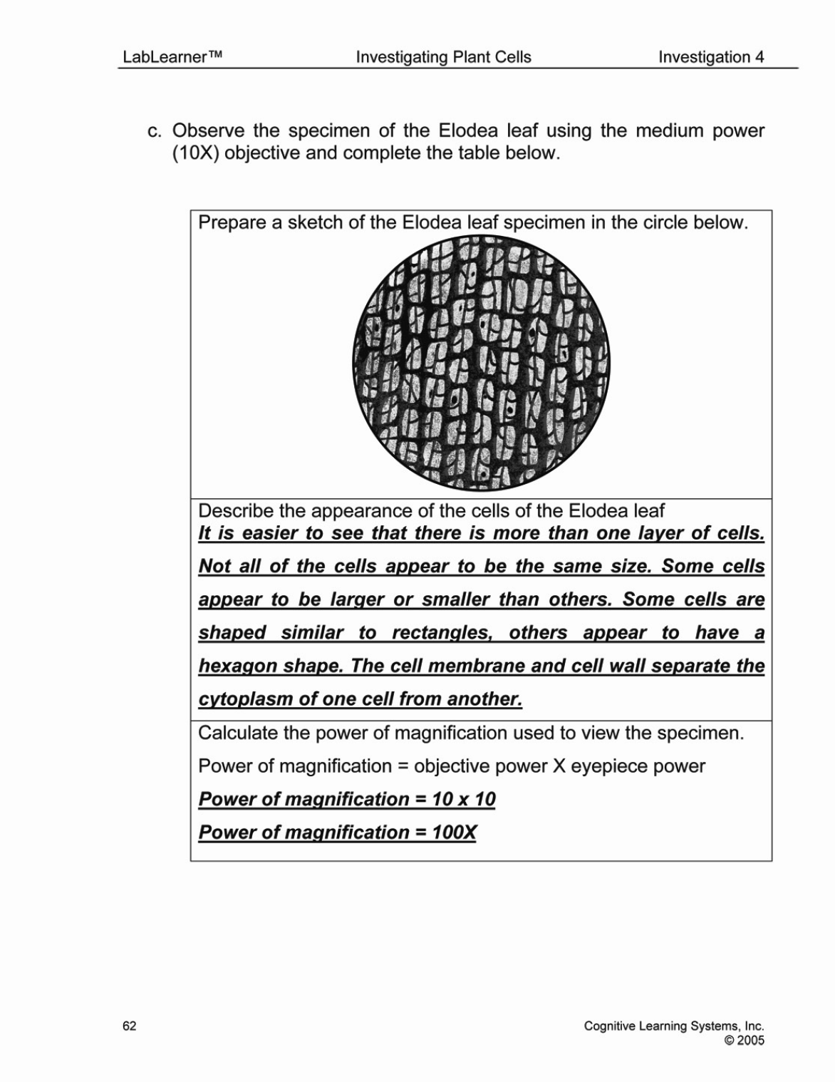
As students study an Elodea leaf and onion specimen they should be aware of the differences in the magnification, resolution, and field of view afforded with each objective lens. They will be asked to apply their observation of these differences as they sketch and describe their observations of the onion cells at each of the different levels of magnification.
As in Investigations One, Two, and Three, students’ observations should help reinforce the concept that lenses refract light and that refraction of light produces images that appear different from the object. Students should also note that regardless of the objective used to view the specimens, the proportions of the cells are maintained when viewing their images.
_____________________________________________
Trial 1
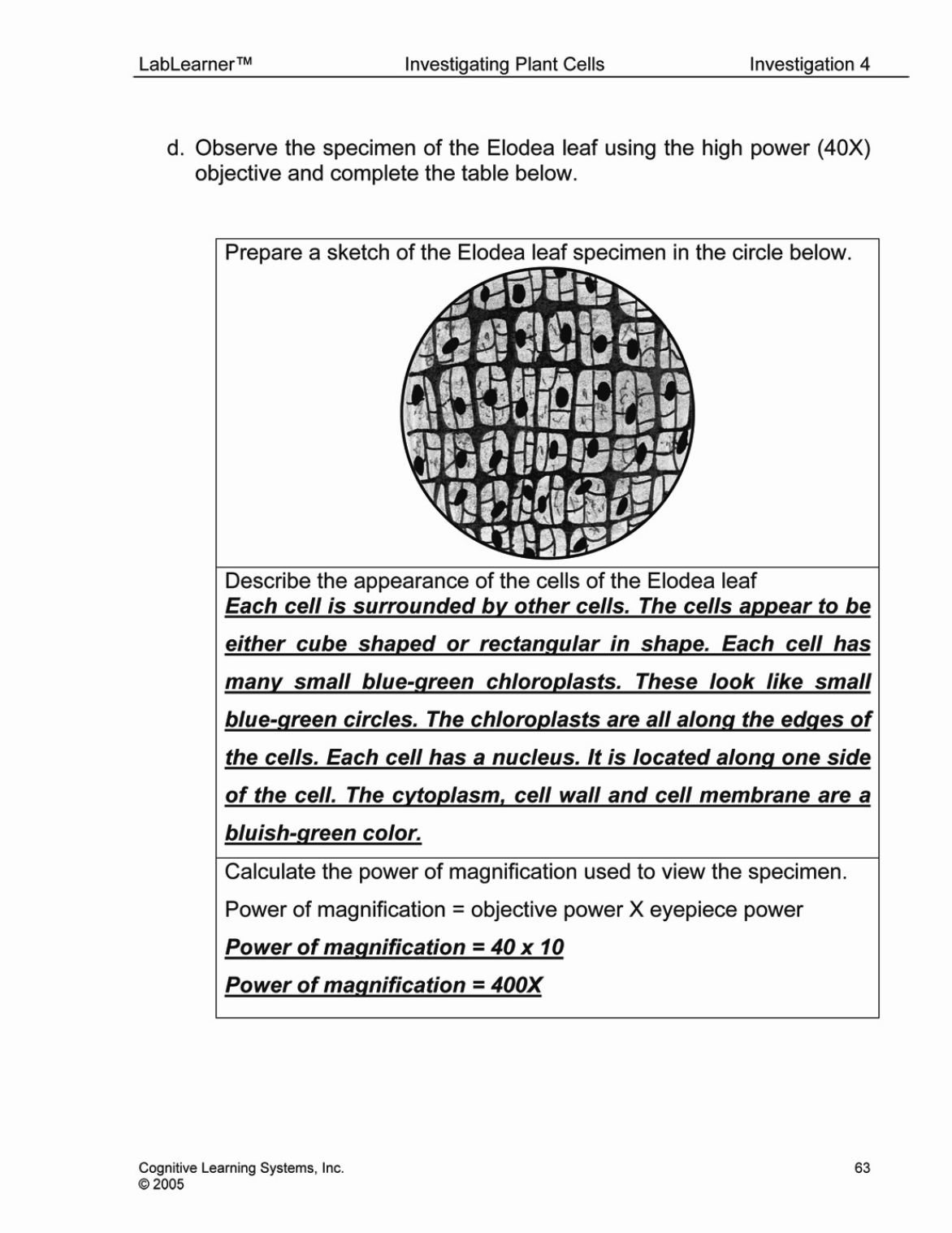
In Trial One, students will have an opportunity to observe organelles and structures contained within the cells of an Elodea leaf. The Elodea leaf is unique in that it is thin enough so that the entire leaf can be mounted on a slide and viewed. As students study the whole mount of the Elodea leaf, they should be able to see the nucleus, cytoplasm, and chloroplasts within each cell in the leaf. Although cells of the Elodea leaf contain both a cell membrane and cell wall, students will not be able to observe the demarcation between these two structures as space between the cell membrane and cell wall of plant cells is so small that it can generally only be observed with microscopes that provide levels of magnification beyond that of a compound microscope, such as the electron microscope. Instead, students will observe a collective boundary of the cell membrane and cell wall between cells of the leaf.
Although each cell of the Elodea leaf does contain a nucleus and multiple chloroplasts, students may have difficulty identifying these organelles. This may occur because the leaf is composed of several layers of cells, one on top of another. It is likely that students will not expect to view a specimen with more than only a cellular layer and therefore will be inexperienced in focusing through the layers. Should this happen, explain the multiple layer nature of the leaf to the students and encourage them to adjust the focus as they view the specimen so that they can observe one layer at a time. This procedure will make it easier for students to identify the nucleus and chloroplasts within each cell.
In addition, some of the Elodea leaf specimens may contain some algae that were retained during the slide preparation If algae are present it may appear as a relatively large, circular organism overlaying some cells. It will be smaller in size than a singular cell, but larger than the nucleus. Students may be tempted to identify algae as the nucleus of each cell because of their circular shape. Help students to differentiate between algae and the nucleus by reminding students that nuclei are found in each cell, whereas algae appear over only a few cells. As students begin this trial, they will be asked the following question:
What information about the structure and function of a leaf and its cells can be learned from the microscopic analysis?
Trial 2
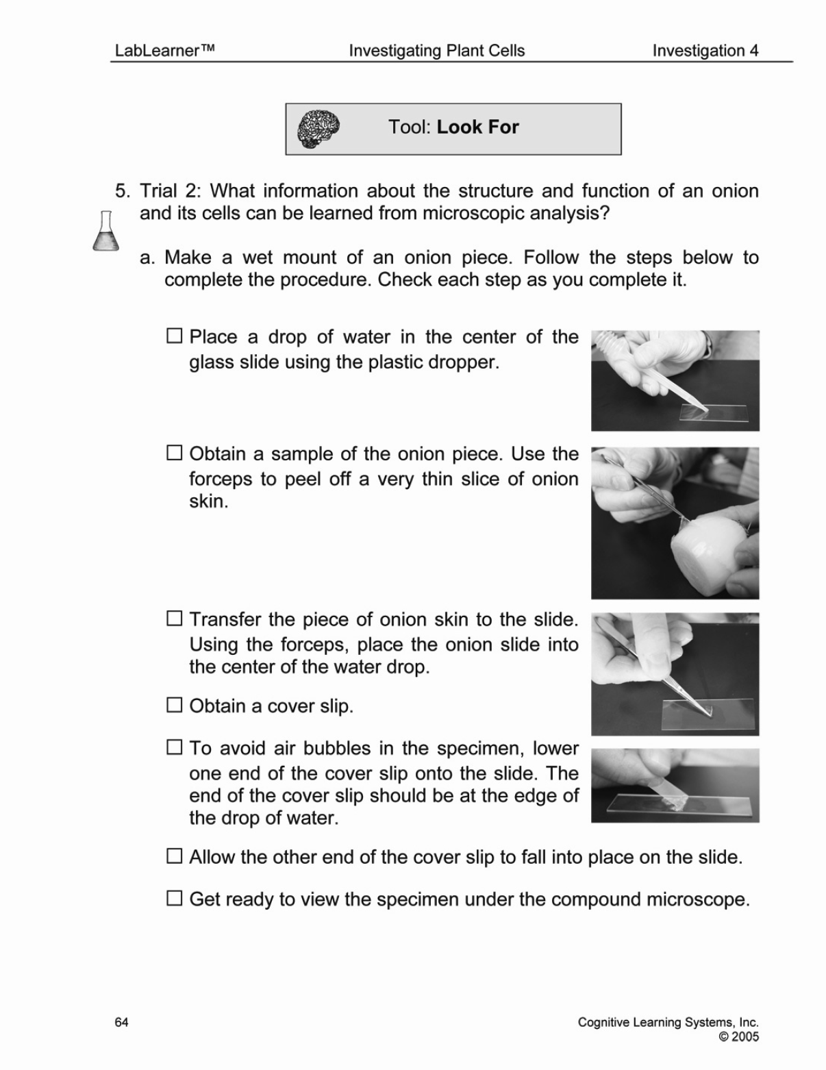
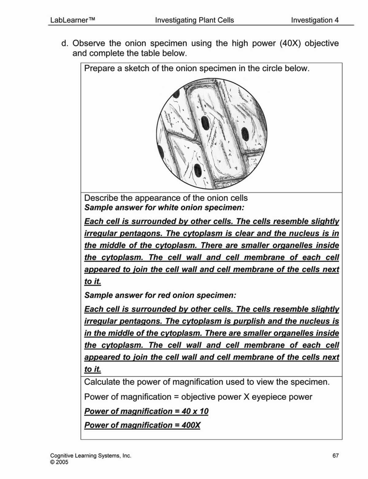
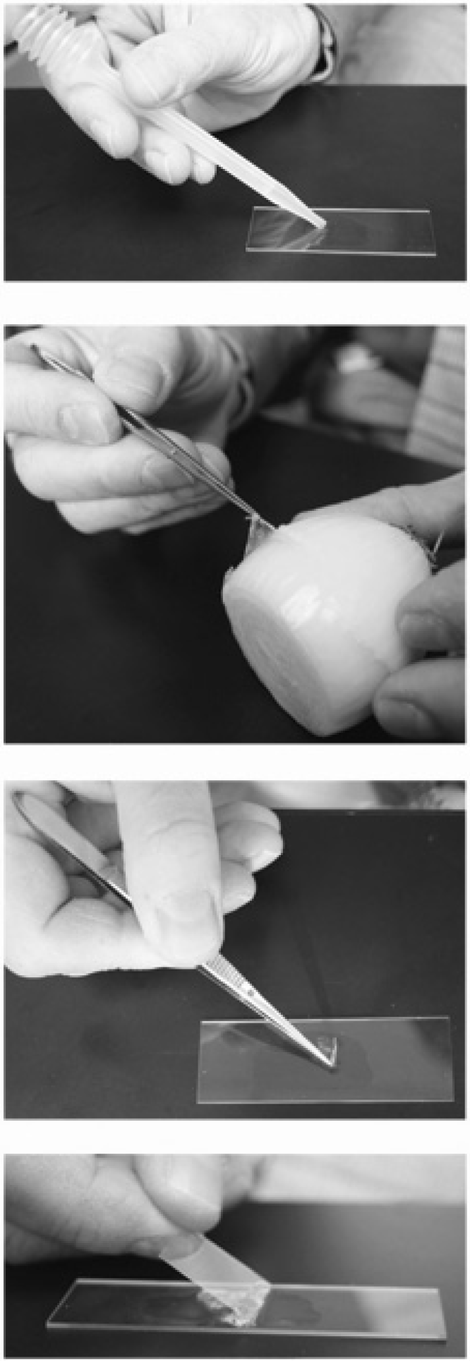 Trial Two has been designed to provide students with a second plant specimen to study and to compare and contrast to the Elodea specimen used in Trial One. In this trial, students will prepare a wet mount of a thin slice of onion and in doing so will have an additional opportunity to practice the tool Wet Mount Slide Preparation from their Procedural Toolbox.
If necessary, review the steps with students prior to beginning this portion of the Lab.
-
- Place a drop of water on the glass slide.
- Gently place the specimen on the water on the slide.
- Place the coverslip on the water on the slide. To minimize air bubbles, place one edge of the coverslip on the slide then tilt the opposite edge until it is resting on the water.
- If the slides contain air bubbles, gently tap on the coverslip. This may effectively rid the slide of air bubbles
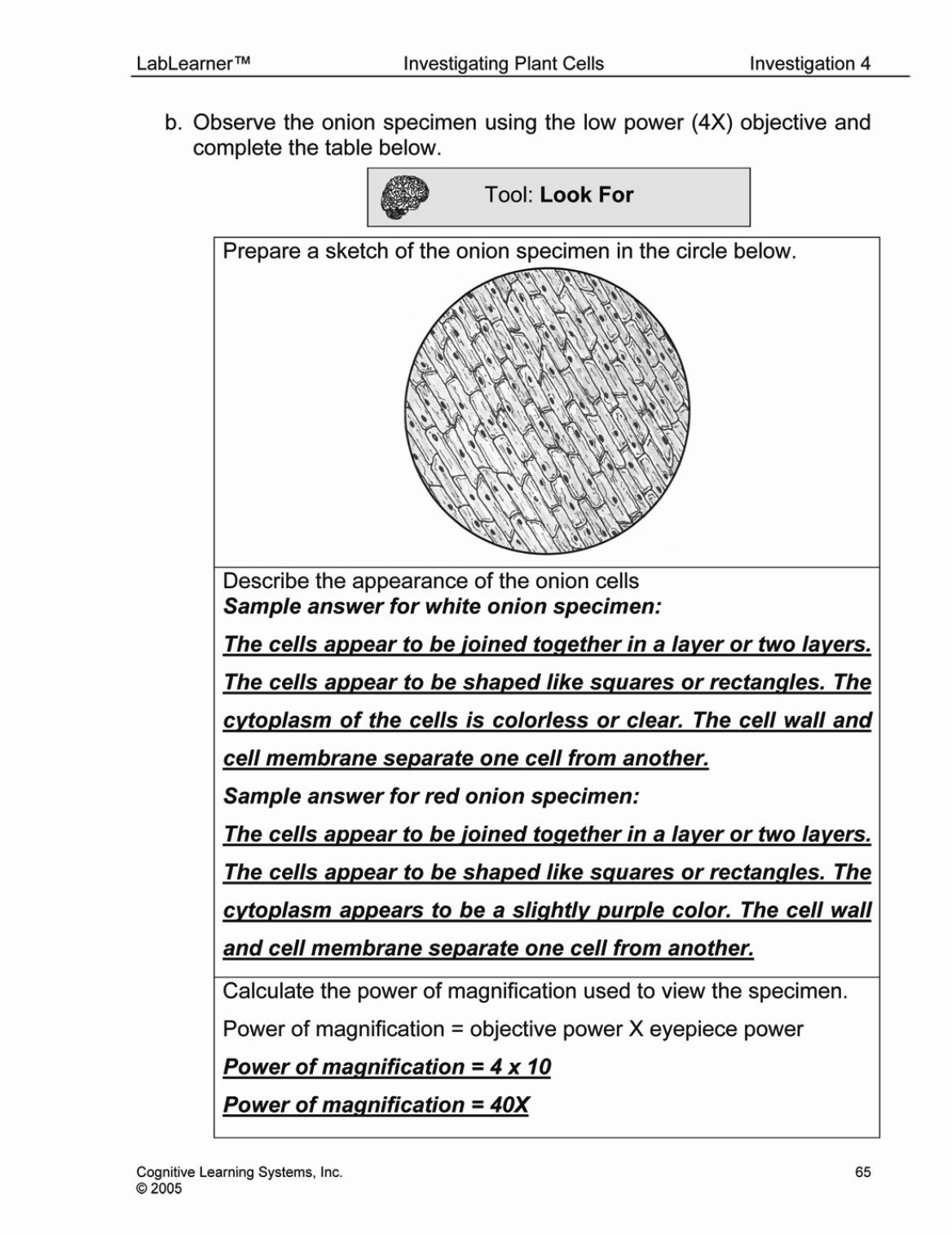
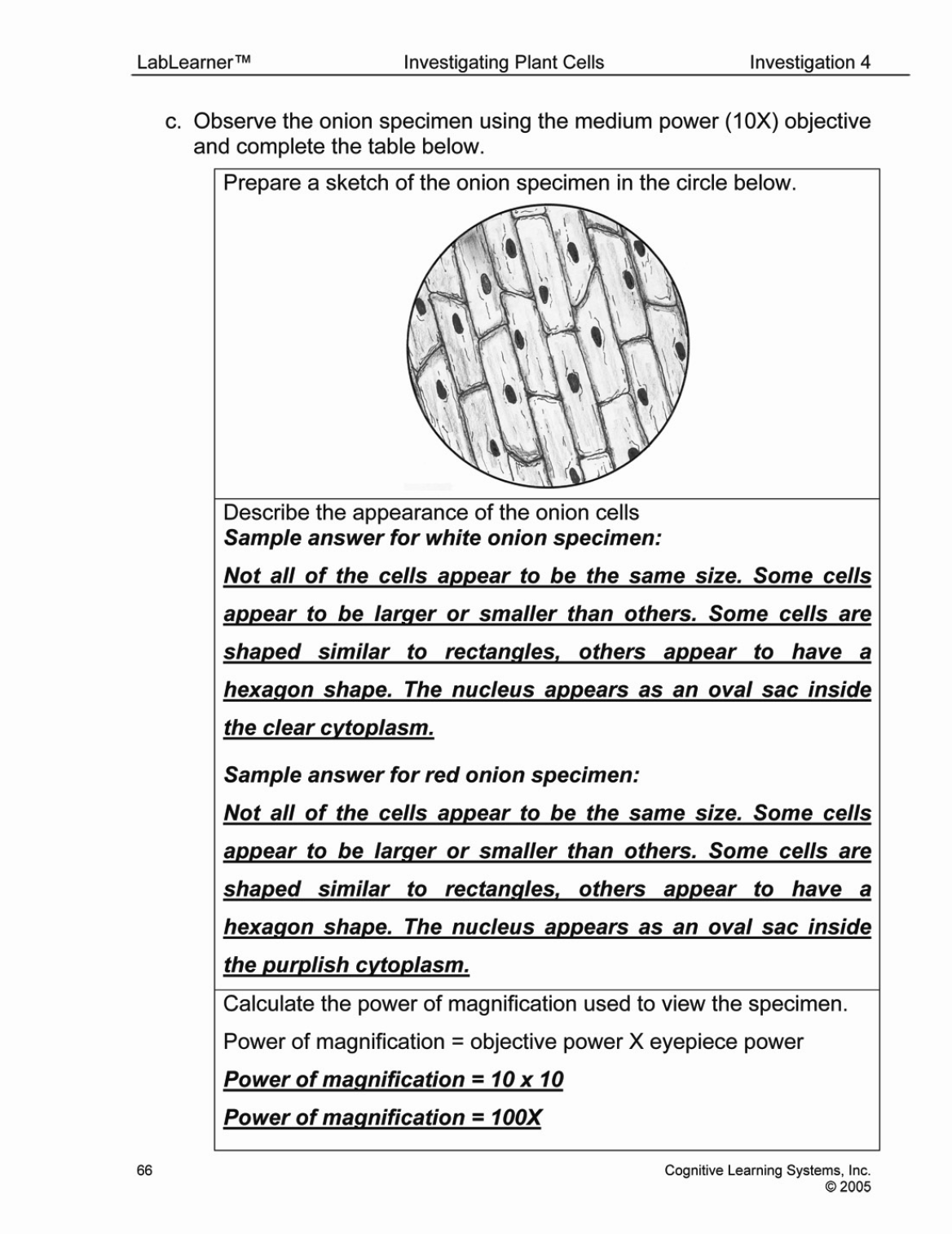
As in Trial 1, students will be asked to identify which parts of the cells they observe including the cell membrane, cytoplasm, cell wall, chloroplast, and nucleus. Although students may not expect to encounter differences in the organelles contained with two different plants they will find that the cells of the onion specimen do not contain chloroplasts. In addition, although the onion cells do contain nuclei, students may have difficulty identifying these organelles. This may occur because of the difficulty in obtaining a thin section of onion skin. It is likely that the students will obtain a piece of onion skin that contains several layers of cells. If this occurs, individual nuclei within the cells will not be easily distinguished. Should this happen, encourage students to adjust their slides in order to view the outer edges of the specimen as it is more likely that these edges contain a singular or a thinner layer of cells.
As students view the onion slice and its cells, they should consider how the arrangement of the organelles affects the function of the cells and the plant. Students will be asked the following question as they complete the trial:
What information about the structure and function of an onion and its cells can be learned from the microscopic analysis?
KEYS: TRIAL 1 and Trial 2

CLEAN UP
Let students know your expectations for clean-up. Ask them to clean up.