
NOTE: The Investigations below are NOT linked in this sample site. They provide a brief overview of Investigation scientific content.
Modeling the Miracle: Tracking Prenatal Development Over Time
Unlike most Units, this activity will span the entire Human Prenatal Development CELL. Each week in the lab, you will use data tables and graphs that provide developmental milestones to follow several parameters, including fetal mass and length, limb and organ development, chances of survival outside the womb, etc.
This experience will condense the 36-week normal human gestation period into four weeks, with model measurements taken at approximately weeks 7, 14, 21, and 28 weeks of development.
Weekly Investigations
Human Prenatal Development: Investigation 1 - Human Chromosomes
In Investigation One, you will learn that humans have 23 pairs of chromosomes. One of these 23 pairs determines the sex of the individual and can be detected by a microscopy technique called a karyotype.
During this Investigation, you will: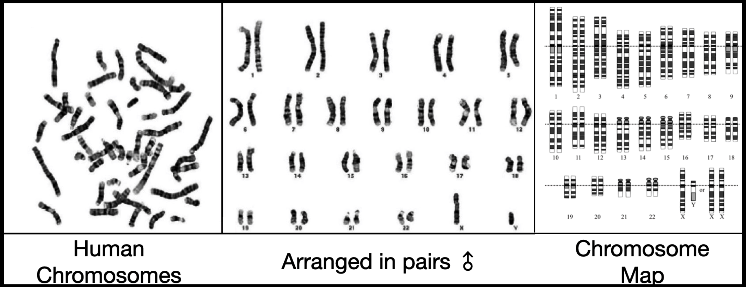
- Examine micrographs of human chromosome karyotypes
- Observe that all but the sex pair of chromosomes appear identical for male and female individuals
- Conclude that while females have two X chromosomes in the sex pair (XX), the male sex pair contains one X and a smaller Y chromosome (XY).
- In lab, students model the process of mitosis to understand every step in this important mechanism of cell division
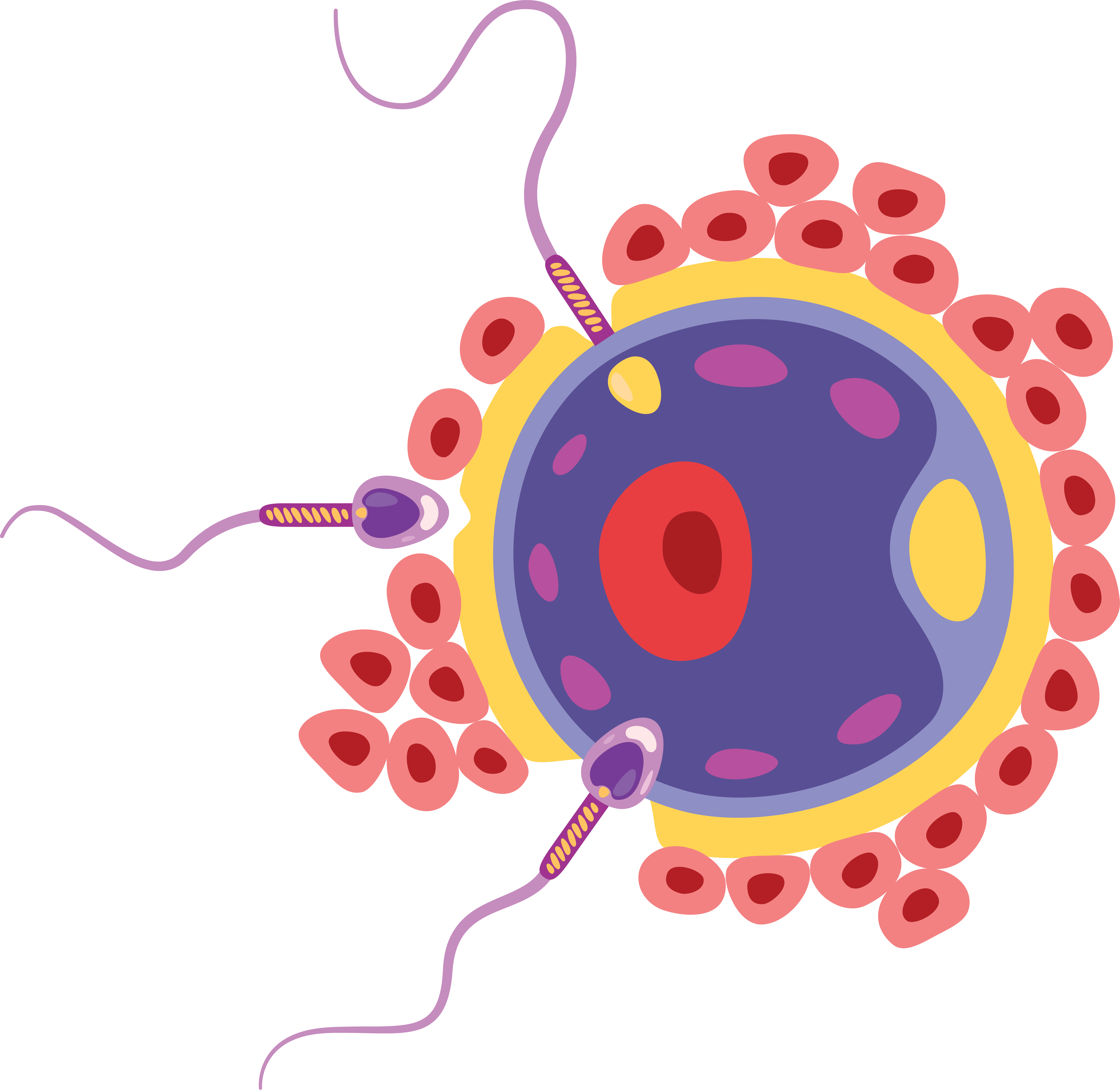
Human Prenatal Development: Investigation 2 - Gamete Formation (Meiosis)
In Investigation Two, you will observe how reduction division (meiosis) creates haploid gametes, containing only half of each of the 23 chromosome pairs.
During this Investigation, you will: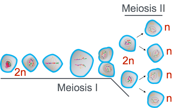
- Learn that while all other body cells (somatic cells) contain 23 pairs of chromosomes (thus 46 chromosomes total), these pairs are separated by reduction division during meiosis.
- The resultant haploid cells contain only one of each original chromosome pair.
- These haploid cells are called gametes; they contain one of each of the 23 human chromosomes and are referred to as spermatozoa (sperm cells) in males and ova (egg cells) in females.
- The male and female gametes combine at fertilization to produce a new individual diploid cell (called a zygote) containing 23 chromosome pairs, which are thus able to perform mitosis and grow rapidly.
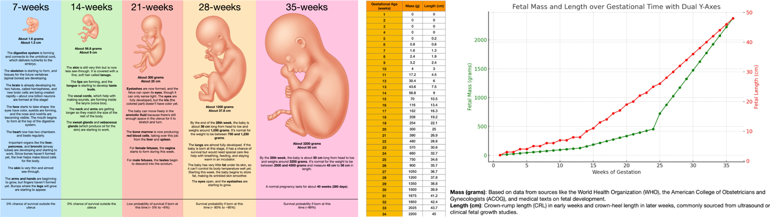
Human Prenatal Development: Investigation 3 - Fetal Development
In Investigation Three, you will continue to follow the initial series of events that occur when male and female gamete cells (spermatozoa and ovum) fuse, forming a zygote. This is followed by a well-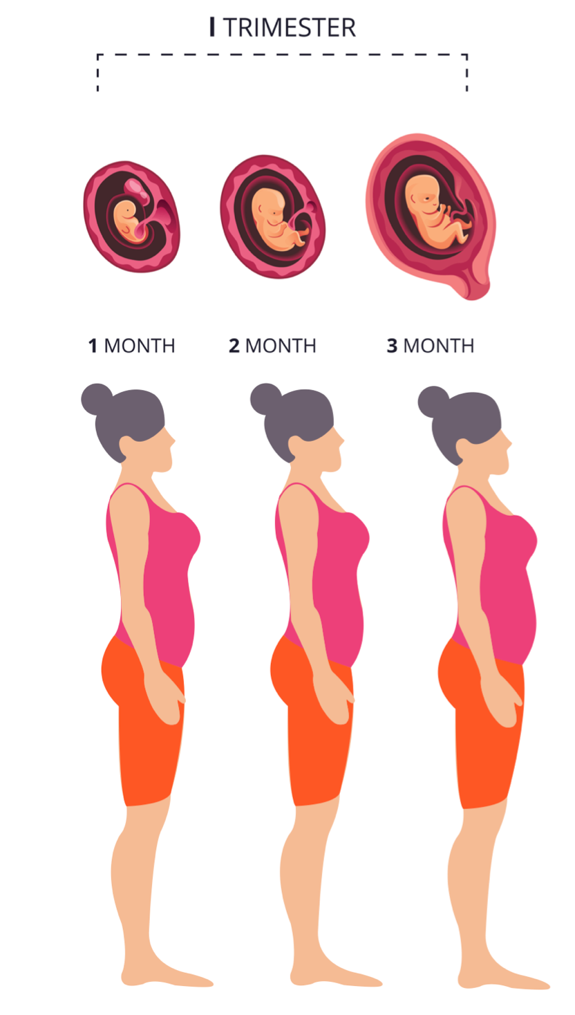 understood sequence of mitotic cell divisions that form a human fetus.
During this Investigation, you will:
- Learn about the defined stages of fetal development.
- Learn how the developing fetus interacts with its mother through a highly specialized tissue called the placenta.
- Learn how toxic molecules are removed from the growing fetus and transported through the placenta to the mother and how nutrients from the mother are transported to the fetus.
- Observe that, by the 21st week of pregnancy, the fetus has grown and is rapidly developing.
Human Prenatal Development: Investigation 4 - Fetal Development and Birth
 In this Investigation, you will follow the developing fetus through the second and third trimester of development.
During this Investigation, you will:
- Follow the development of fetal organ systems and dissect a fetal pig to examine the extent of organ development.
- Learn that the baby may survive premature birth as early as the end of the sixth month of pregnancy.
- examine life-size anatomical models of a full-term pregnancy.
- Learn how ultrasound works and how it permits physicians to follow fetal development.
