Teacher Portal:
Genes and Proteins
Investigation 2 – Concept Day
ZERO-IN

Italicized font represents information to be shared orally or physically completed with the students at this time.
The non-italicized font represents additional information included to support the teacher’s understanding of the content being introduced within the CELL.
ASK WHY
Remind students that genes are instructions, which tell your body how to make all the proteins it needs to survive and grow. By identifying each of these proteins, scientists hope to better understand how your body works, and what is happening when it doesn’t work properly. They hope this knowledge will eventually lead to more effective medicines and treatments.
BRANCH OUT
Explain to students that a statistical geneticist develops software to perform genetic analysis, for example, quantitative trait loci mapping.
PRINT IT
Use your browser to download a printable PDF as a help during the slide presentation and to make additional notes. In your browser, go to File > Print and then choose to save as PDF.
NAVIGATE IT
Once the slide presentation is launched
- use your left and right arrows to advance or go back in the slide presentation, and
- hover your mouse over the left edge of the presentation to get a view of the thumbnails for all the slides so that you can quickly move anywhere in the presentation.
- Click HERE to launch the slide presentation for the CELL.
SHARE IT
SLIDE GENES-2-1
In this Investigation, we pick up on the topic of protein synthesis (protein translation). More specifically, most of this presentation will be devoted to helping students understand the importance of proteins in cells and how a protein’s function is directly dependent on the DNA sequence that codes for its synthesis. We will finish with a discussion of the importance of mutations as related to protein structure and human disease.
______________________________________________
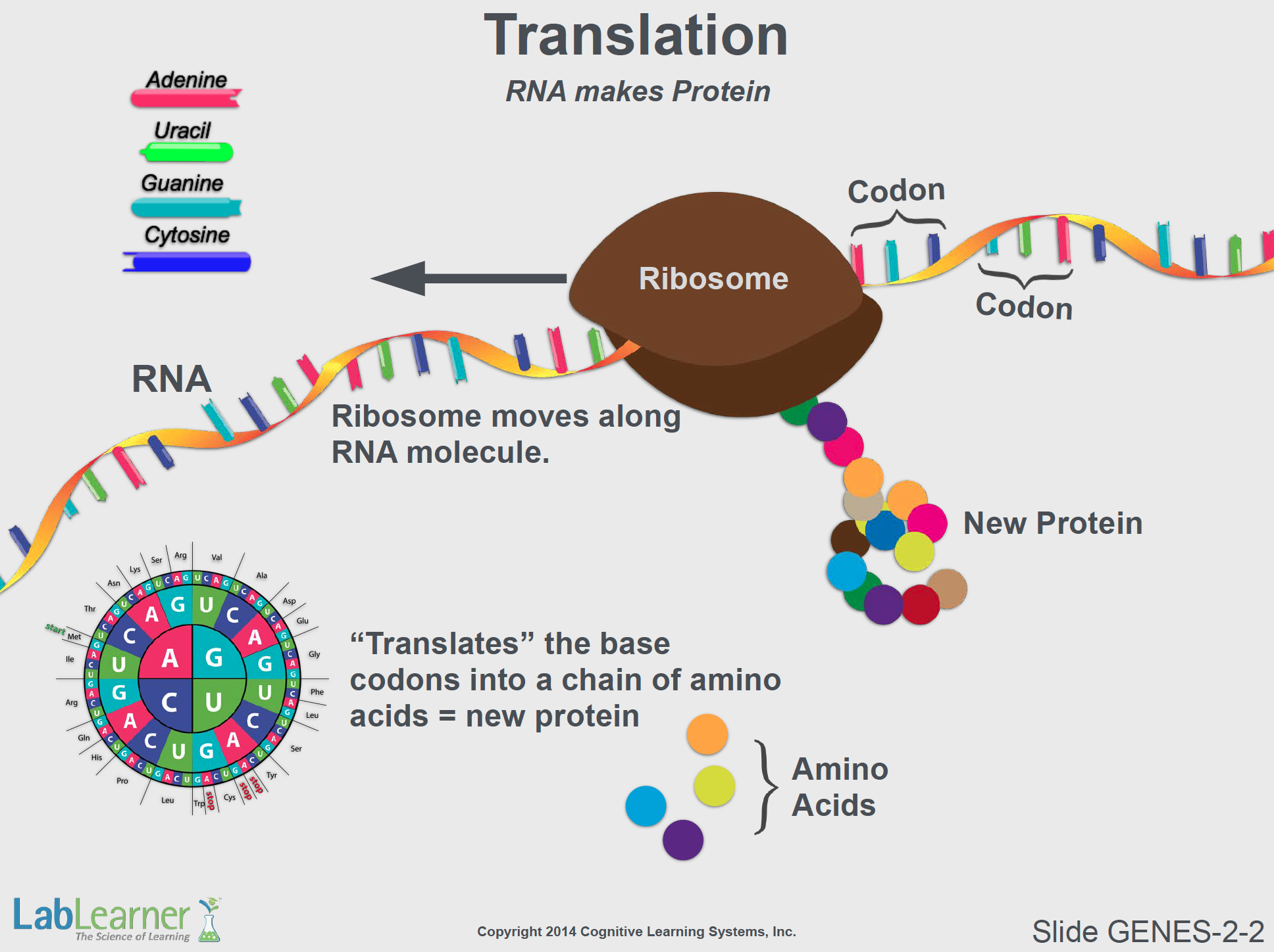
SLIDE GENES-2-2
- This slide was originally presented in the previous Investigation in terms of the role of protein synthesis in the Central Dogma of Molecular Biology (DNA makes RNA makes protein). It serves as a good start off for a discussion of protein structure and function.
- While only the RNA strand is shown here, students should recall that this RNA molecule (also called mRNA for messenger RNA), was transcribed directly from the original DNA template in the cell nucleus. Thus, the properties of all proteins in our bodies are dictated by their DNA sequences and inherited from our parents.
- In this slide, the essential point to highlight is that the sequence of bases on the mRNA molecule is decoded by cytoplasmic ribosomes by reading a series of three-base codons. Each codon encodes for either a specific amino acid that will be added to the growing protein chain or will start or stop protein translation (start and stop codons, respectively).
Note: We must never forget that, regardless of size or complexity, a protein molecule is still a molecule. It is composed of atoms that interact or bind to each other. A protein’s total mass therefore is equal to the sum total of all of the individual atoms that it is composed of. Consequently, while a simple sugar like glucose (C6H12O6) contains only 24 atoms (6 carbon, 12 hydrogen, and 6 oxygen) and a protein molecule may contain well in access of hundreds of thousands of atoms, both are nonetheless molecules.
______________________________________________
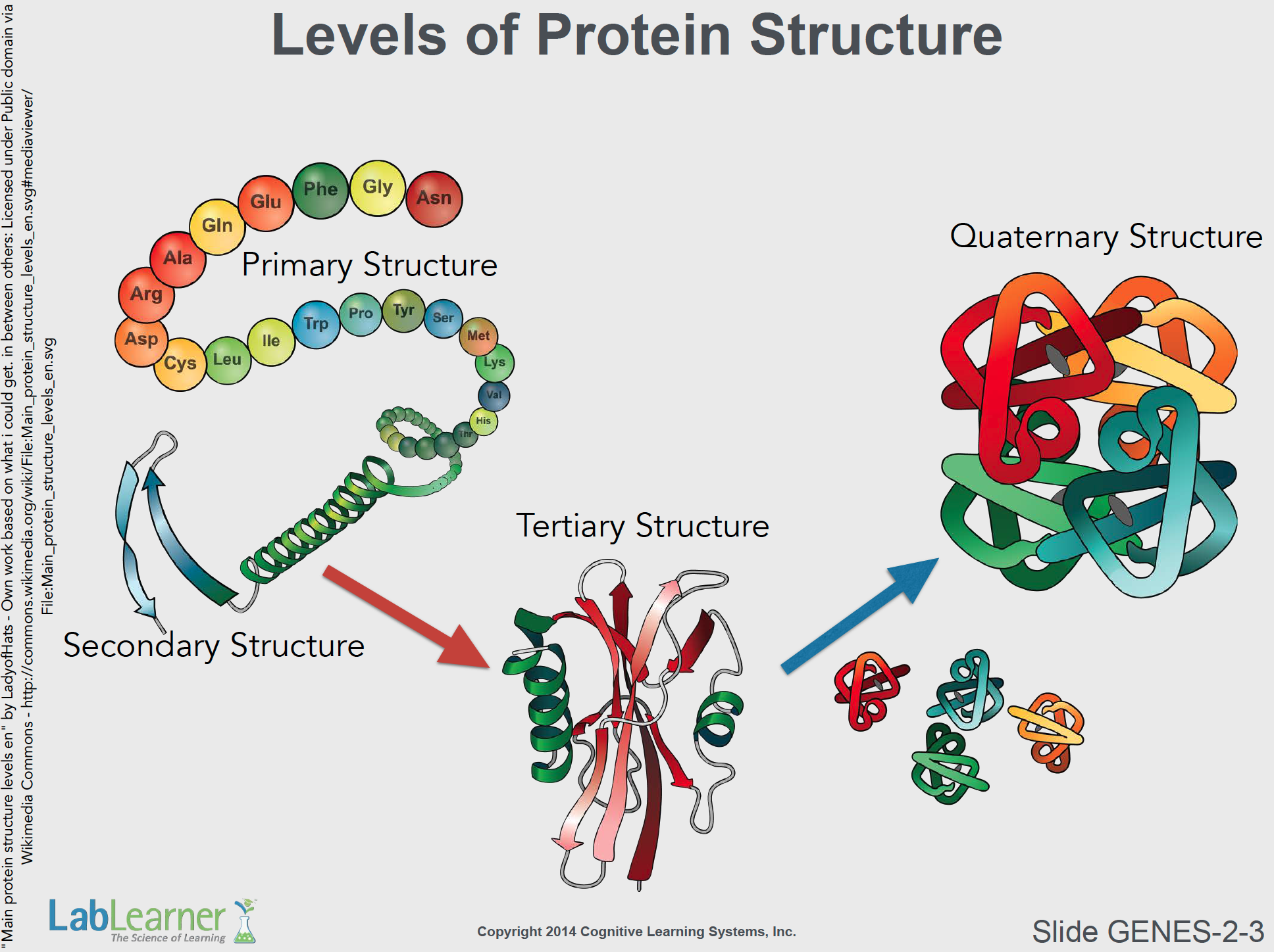
SLIDE GENES-2-3
- This important slide depicts the varying levels of structure within an individual protein molecule. The levels are referred to as primary (1o), secondary (2o), tertiary (3o), and quaternary (4o). At this point, students needn’t be concerned about the details of protein structure other than to know that a protein’s component amino acids and overall shape determine its function. Thus, anything that affects a protein’s shape/structure can alter its function. Mutations in the DNA sequence of a protein gene, for example, can alter the protein’s function. Other factors such as temperature, salt concentration, and other conditions can also alter protein function but will not be discussed here.
- Primary Structure: This refers simply to the sequence of amino acids in a protein. This sequence, of course, is dictated by the DNA sequence of the gene. This amino acid sequence is sometimes referred to as a polypeptide chain. This is because the bonds that hold the amino acids together in a linear chain are call peptide bonds.
- Secondary Structure: Due to the physical properties of the individual amino acids (remember, there are 20 different amino acids), sections of the polypeptide chain may interact with each other to form local structures. For example, a stretch of amino acids may interact with each other for a spiraling helix. They may also for zigzag structures and flat sheets. Such localized secondary structures may be important for a protein’s function.
- Tertiary Structure: A completed protein may have many secondary structures and amino acid sequences that attract and interact with each other. Consequently, the overall amino acid chain often folds in upon itself in a very specific manner to form a tertiary structure. One important result of such tertiary structure is that the folded protein’s three-dimensional structure may form surface areas that recognize and bind to other molecules. Such a structure would clearly be important for the protein’s function. Oftentimes, mutations that affect protein function do so because a replaced amino acid, caused by the mutation, does not lead to the formation of functional binding sites on the protein molecule.
- Quaternary Structure: Finally, many proteins are actually composed of more than one polypeptide chain. In such cases, two or more different polypeptide chains, each with their own tertiary structure, bind to each other in very specific arrangements. When this happens, the individual polypeptide chains within the final protein molecule are typically referred to as subunits of the protein.
- In the next slide, we will look at the protein hemoglobin. It is a protein that has two each of two different polypeptide chain subunits and thus provides an example of a protein with a quaternary structure.
______________________________________________

SLIDE GENES-2-4
- This slide shows the structure of the human hemoglobin molecule. Hemoglobin is a protein found in red blood cells and is actually what makes our blood red. It is composed of two alpha and two beta subunits. Each of the subunits contains an iron group (a heme group) that can bind a molecule of oxygen (the heme group is shown in green in this model).
- This essential protein is made in incredibly large amounts in red blood cells and is responsible for carrying oxygen from the lung to the tissues of the body. We will see later, the result of a single mutation in the hemoglobin gene.
______________________________________________
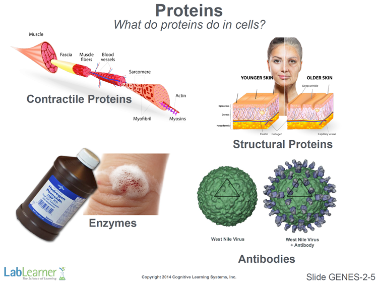
SLIDE GENES-2-5
- We now turn to a discussion of the function of the protein in cells. This is simply an introductory slide. Proteins perform many different functions in cells and they make many thousands of different proteins. A simple way to look at it is to assume that, even if we do not understand the function of a particular protein in a cell, the very fact that it is made means that it is important, if not essential, to the cell’s survival.
- As shown on this slide, the examples of protein function we will discuss are contractile proteins, structural proteins, enzymes, and antibodies.
______________________________________________
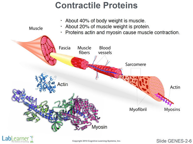
SLIDE GENES-2-6
- Two major contractile proteins are actin and myosin. It is the interaction between actin and myosin that causes muscles to contract and exert force. In muscle tissue and in other cells, the actin protein interacts with itself to form long fibers. This contributes to the characteristics of muscle fibers. The energy molecule ATP is required for the interaction between the action and myosin molecules to generate the force of muscle contraction.
______________________________________________
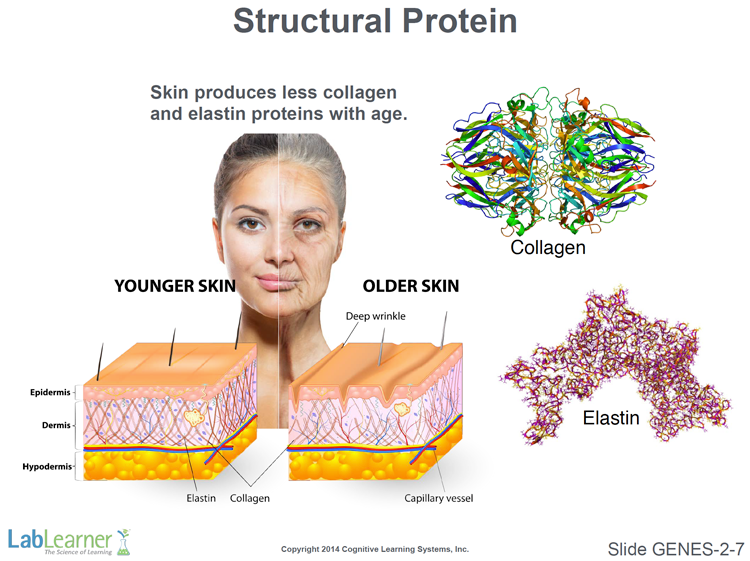
SLIDE GENES-2-7
- Collagen and elastin are two structural proteins found in the dermis layer of skin and other tissues that require strong structural support, such as tendons and ligaments. The collagen protein molecule forms elongated fibers and along with the protein elastin, helps support the shape and flexibility of the skin.
- Unfortunately, with advancing age, the amount of these proteins made by cells in the skin decreases, and the structures that they form in the dermis break down. This results in the thinning and reduction of firmness of the skin that produces wrinkles.
______________________________________________
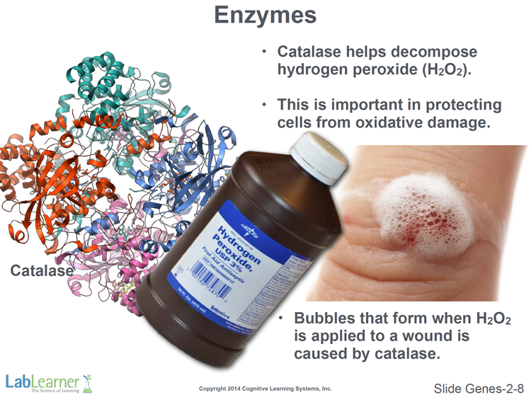
SLIDE GENES-2-8
- Enzymes are protein molecules that act as catalysts in chemical reactions. That is, they promote or speed up chemical reactions without being consumed in the reactions. They are neither reagents nor products; they simply help transform chemical reagents into products.
- The enzyme highlighted on this slide is catalase. It is found in all organisms that live in an environment that contains oxygen. Although essential for life, oxygen also cause various kinds of damage to cells as a result of a number of chemical reactions involved in metabolism. One of these is the formation of the toxic substance hydrogen peroxide (H2O2). The enzyme catalase helps convert hydrogen peroxide to water and oxygen:

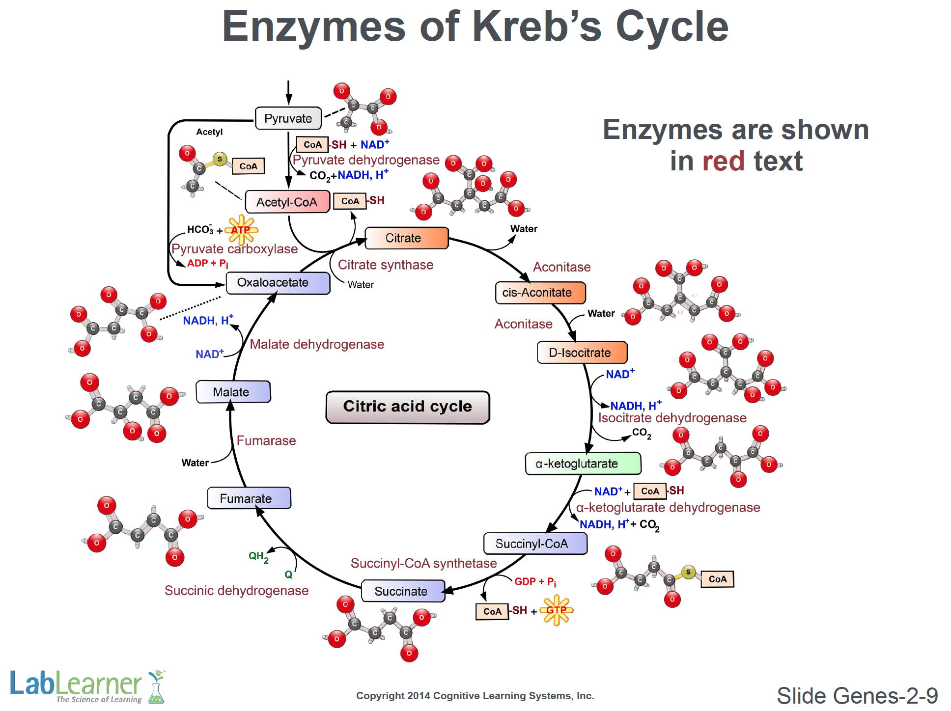
- There are thousands of different enzymes in cells, all performing various essential functions in the overall chemistry of the organism. Enzymes often pass the product of one chemical reaction on to another enzyme that will perform the next catalytic step in a long series of chemical reactions. One such series of chemical reactions is referred to as the Kreb’s Cycle and is shown on the next slide.
______________________________________________
SLIDE GENES-2-9
Note: This slide is not meant to intimidate or frighten students (or teachers). No details whatsoever of this important cycle of reactions need be considered at this point. It is simply included here to impress upon students the vast number and importance of enzymes in cells and to begin to communicate to them the wonderful chemical complexity of life.
- There are many thousands of different enzymes in cells. Therefore, the complex series of enzyme reactions of the Kreb’s Cycle probable about 1/1,000th of the enzymatic reactions that actually occur in cells!
______________________________________________
SLIDE GENES-2-10
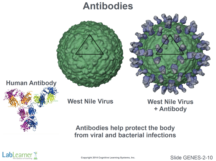
- The final type of protein we will discuss are the antibodies. Antibodies are made by our immune system and are used to combat infections by recognizing and binding to foreign invaders such as bacteria and viruses.
- The polypeptide chains of antibodies are synthesized to recognize and bind to specific proteins on the surface of the invaders. This then targets the bacteria of viruses for destruction by the cells of the immune system.
______________________________________________
SLIDE GENES-2-11
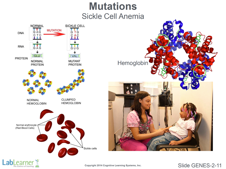
- We finish this Investigation with two examples of mutations that cause disease in humans. In this slide, we discuss the disease Sickle Cell Anemia.
- Here again, we see the human hemoglobin protein molecule on the upper right. A single point mutation in the gene that encodes the beta subunit of hemoglobin causes this disease. As shown in the upper left of this slide, the normal DNA sequence of GAG is mutated to GTG, which is an adenine base is replaced, through mutation, with a thymine base. After transcription, the resulting mRNA molecule has a mutant codon GUG rather than the normal (or wild type) GAG. Therefore, instead of inserting the amino acid Glu (Glutamic acid) into the growing beta hemoglobin chain, the ribosome inserts the amino acid Val (Valine). That’s all it takes to cause this disease.
- The mutant hemoglobin molecule causes normally smooth, disc-shaped red blood cells to form irregular, sickle-shaped blood cells. This causes them to get trapped and destroyed in the spleen and causes other problems throughout the circulatory system.
- Since the root of the disease is a mutation in the DNA sequence, this is a heritable disease – it is passed from one generation to the next. According to the CDC (Center for Disease Control), sickle cell disease occurs among about 1 out of every 500 African-American births and 1 out of every 36,000 Hispanic-American births.
______________________________________________
SLIDE GENES-2-12
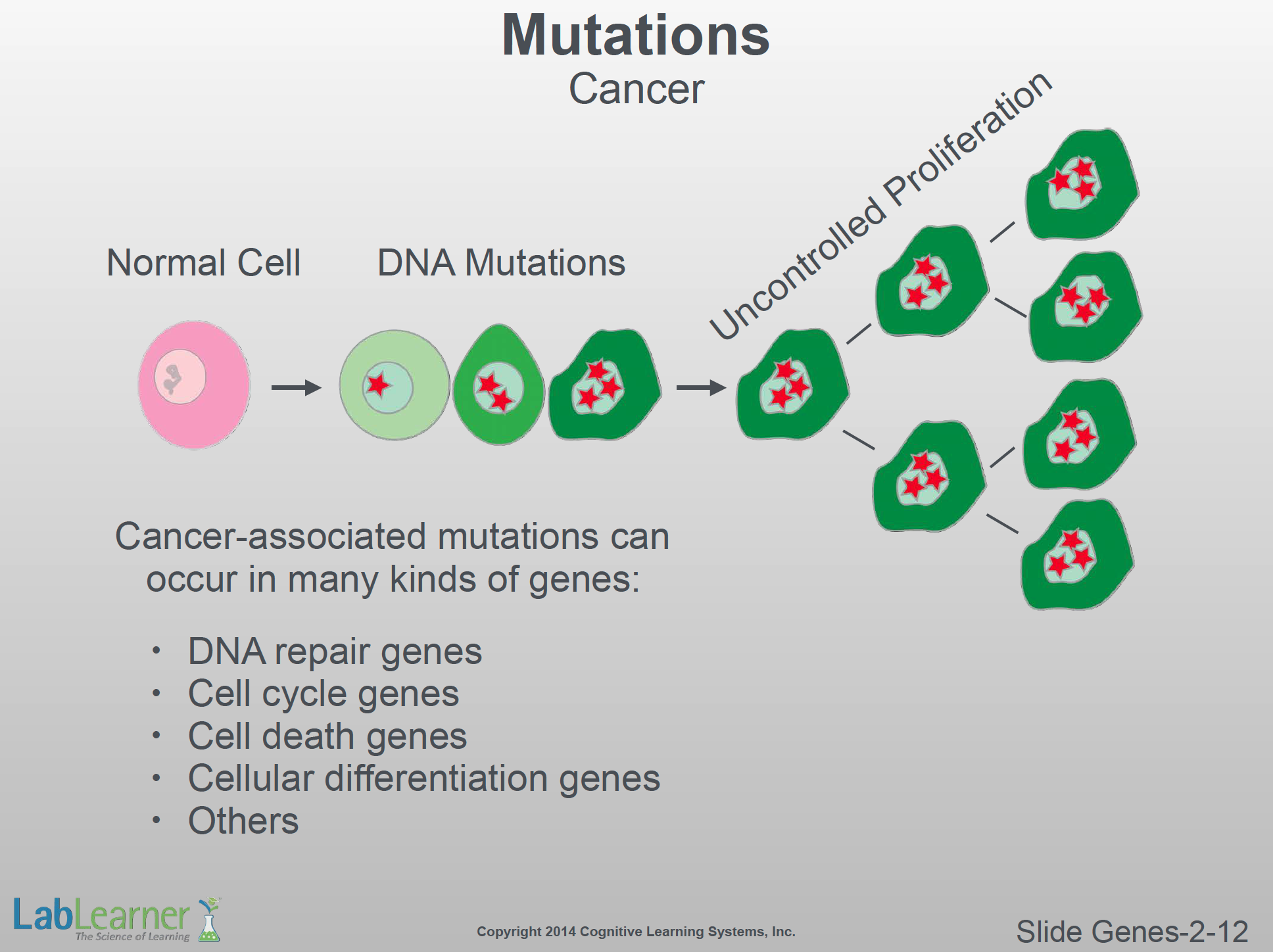
- Some DNA mutations are also responsible for causing cancer. Not only can DNA mutations be caused by environmental factors such as UV irradiation and toxins, but they may also be permanent features of the DNA inherited from our parents. Thus, certain cancers are now known to be hereditary. We can inherit a predisposition for certain kinds of cancer.
- This slide shows how a normal cell, through a process of several mutations, becomes cancerous. Once mutations accumulate in a cell, it may lose the ability to perform its normal functions. Perhaps worse, its ability to control its own cell division and proliferation may be impaired. The resultant uncontrolled cell proliferation can cause masses and tumors.
- Finally, some of the types of cancer-associated mutations that may occur are shown on the lower left of this slide. In the next CELL, Cell Cycle and Cancer, we will discuss this topic in much greater detail.
______________________________________________