Teacher Portal:
Cellular Organization
Investigation 3 – Lab
ZERO-IN
Italicized font represents information to be shared orally or physically completed with the students at this time.
The non-italicized font represents additional information included to support the teacher’s understanding of the content being introduced within the CELL.
MINDSET
This Investigation is designed to:
- reinforce microscope operation and preparation of a wet mount slide,
- aid students in identifying common structures of plant cells including chloroplasts, cell walls, cell membranes and nuclei,
- aid students in comparing structures and organization of plant cells and tissues to those of animal cells and tissues,
- help students understand that although generalizations about cellular structures of plant and animal cells can be made, all cells from the same organism may not have identical cellular structures or organelles, and
- help students understand that the presence or absence of some cellular structures governs cellular and tissue function
BE PREPARED
Teacher Preparation for the Investigation includes the following. This preparation should be done prior to students arriving in the lab.
- Remove and discard the outer brown layer of the onion. Cut the onion into five wedges so that each student group has one small wedge.
- Place the bottles of immersion oil next to each microscope.
- Place the remaining materials at the distribution center.
- Divide the class into five cooperative groups.
Note: Each student lab group will need the materials listed below.
Student Preparation for the Investigation includes having students gather the following materials. This preparation takes place on lab day after student lab groups have settled at their assigned lab tables.
Note: The materials are listed in students’ SDRs. They are also listed below for your reference.
- (1) microscope
- (1) plastic dropper
- (1) forceps
- (1) glass slide
- (1) coverslip
- (1) paper towel
- (1) section of onion
- (1) human cheek cell slide
- (1) human blood smear slide
- (1) Elodea leaf slide
- (1) large intestine slide (human colon)
- (1) Elodea stem slide
- (1) sheet of lens tissue
- (1) 100 ml beaker with 50 ml of water.
Direct one student from each lab group to collect the materials listed in their SDRs.
INVESTIGATE
- Informally assess students’ knowledge of the parts of an animal cell and a plant cell. Include the cell wall, cell membrane, cytoplasm, nucleus, and chloroplasts. Sketch an animal cell and a plant cell on the board if necessary. Students should understand that plant cells have cell walls and chloroplasts, while animal cells do not.
- Encourage students to reflect on the PreLab video as they move through the procedural steps.
- Explain to students that during the Experiment, every procedural step is important. If one step is skipped, data can become invalid. To help students keep on track, direct them to read each step thoroughly, complete the step, then check it off (Read it – Do it – Check it off).
- Direct students to complete the procedural steps in their SDRs.
Note: The procedural steps are listed below for your reference. Teacher “Notes” are inserted, as needed, to help facilitate the lab.
- Peel a very thin layer of onion off your section. The sample must be very thin so that it is only one layer of cells.
- Create a wet mount slide of an onion. Refer to the procedure to the procedure, Wet Mount Slide Preparation.
- Observe the onion slide under the microscope. Refer to the procedure, Microscope Use and Operation, if you need help. You may be able to more easily view the cells near the edge of your sample. The edges are likely to be thinner than the center of the specimen.
Note: Do not use the 100X oil immersion objective with wet mount slides. You may use the 100X objective with prepared slides, because they are sealed so the oil cannot go under the coverslip.
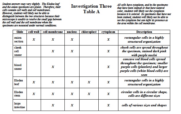
- Look for the cell structures listed in Table A.
- Record: As you see and identify the structures, place a check in the appropriate column of Table A.
- Record: Write any general observations about the slide in the last column of Table A.
- Observe each of the other prepared slides listed in Table A under the microscope.
- Record: Identify the structures and write your observations for each slide in Table A.
CLEAN UP
Let students know your expectations for clean up. Ask them to clean up.