Teacher Portal:
Cell Cycle and Cancer
Investigation 3 – Lab

ZERO-IN
Italicized font represents information to be shared orally or physically completed with the students at this time.
The non-italicized font represents additional information included to support the teacher’s understanding of the content being introduced within the CELL.
MINDSET
This Investigation is designed to:
- demonstrate the relationship between control of the cell cycle and cancer,
- allow an opportunity for students to examine and compare tissue sections of normal and cancerous tissues,
- provide an opportunity to model and investigate the changes in cellular organization that can occur with the development of cancer,
- demonstrate how changes in cellular organization can affect the function of a tissue or organ, and
- demonstrate the loss of regulation of the cell cycle during cancer via that cell cycle model from Investigation Two.
BE PREPARED
Teacher Preparation for the Investigation includes the following. This preparation should be done prior to students arriving in the lab.
- Cut and place plastic tubing, marbles, push pins, packing tape and permanent markers at a central distribution center.
- Place one microscope at each group station.
- Place the onion root tip slide and one bottle of immersion oil at each group station.
- Divide students into five cooperative groups.
Note: There is no Student Preparation for Investigation 3 Lab.
INVESTIGATE
- Encourage students to reflect on the PreLab video as they move through the procedural steps.
- Explain to students that during the Experiment, every procedural step is important. If one step is skipped, data can become invalid. To help students keep on track, direct them to read each step thoroughly, complete the step, then check it off (Read it – Do it – Check it off).
- Direct students to complete the procedural steps in their SDRs.
Note: The procedural steps are listed below for your reference. Teacher “Notes” are inserted, as needed, to help facilitate the lab.
Trial 1:
Note: In this trial, students will create a model of a tissue in which contact inhibition prevents cells from dividing uncontrollably. Then, they will model what would happen if the tissue became cancerous,
- In this trial, you will examine the role of contact inhibition in preventing cells from continuously dividing.
A. Separate the gram cubes into two piles by color.
B. Check that you have a pile of 9 cubes of the same color. These represent normal cells.
C. Check that you have a pile of 81 cubes of the same color but a different color from the first pile. These represent cancerous cells.
D. Place one of the gram cubes from the smaller pile into the 100 ml beaker. This represents tissue with “normal” cells in it.
2. A normal cell is a cell with a normally regulated cell cycle. In this model, a normal cell will divide once every 15 seconds. Assume that only 1 normal cell of any group divides every 15 seconds. The remaining cells are in the G1 (GAP1) phase of the cell cycle and are not currently dividing.
3. Start the timer. Add 2-gram cubes of the same color every 15 seconds for 1 minute.
- Look at the cubes in the beaker. Imagine that these normal cells are regulated by contact inhibition. Why did they stop dividing after 1 minute? The cells stop dividing because they came in contact with other cells surrounding them (the cubes fill the bottom of the beaker). When the cells interacted with neighboring cells, the cell cycle regulatory proteins would have been produced and would have stopped the cell cycle, thus inhibiting cell division.
- In this Trial, you will model what happens when one of the normal cells is transformed into a cancerous cell.
- Remove one of your normal cells from the beaker and replace it with a gram cube of another color. This represents a cancerous cell. You now have a beaker with many normal cells and 1 “cancerous” cell.
- Organize the large pile of remaining gram cubes into smaller piles.
A. Create a pile of 1 cubes.
B. Create a pile of 2 cubes.
C. Create a pile of 4 cubes.
D. Create a pile of 8 cubes.
E. Create a pile of 16 cubes.
7. In this model, every cancer cell in the beaker will divide once every 15 seconds. Use the table to discuss with your group why the total number of gram cubes to be added is as it is shown.
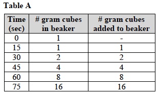
- Reset the timer then begin timing for 1 minute. After every 15 seconds add two cancerous gram cubes for each cancerous cell in the beaker using Table A as a guide.
- At the end of 75 seconds, what happened to the gram cubes you were adding to the beaker? Was contact inhibition effective in preventing cell division? The gram cubes were piled on top of each other to the point that they had spilled out of the beaker. Contact inhibition was not effective in preventing cell division.
- How is the organization of the cells in the cancerous “tissue” different from the normal “tissue” you had made in number 3 above? Remember, both “tissues” grew for 1 minute. The normal tissue had a single layer of cubes (cells) on the bottom of the beaker after 1 minute. However, after 1 minute the cancerous tissue had cells piled up on top of each other to the point where they spilled out of the beaker.
Trial 2:
- Set up the microscope according to the Procedure, Microscope Use and Operation.
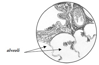
- Review the normal lung slide. Place the slide on the microscope stage and focus on the lowest power. Then, focus on each of the higher power objectives up to 40X. Do not use the 100X objective.
- Record: Write or draw your observations while examining a field containing alveoli with the 40X objective in the space below:
- Place the lung adenocarcinoma slide on the microscope stage. Focus first on the lowest power, and then focus on each of the higher power objectives up to 40X. Do not use the 100X objective.
- Record: Look at the different cell types and the organization of the cells by moving the microscope stage as needed. Draw a picture of a field containing alveoli as seen with the 40X objective.
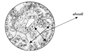
- Compare the normal and the lung cancer slide. What are the differences in cellular organization in the adenocarcinoma and normal lung tissues you examined? Where will the inhaled air go in the tissue containing cancer cells? The normal lung slide shows spaces in the alveoli for the air to pass through. However, the air will not be able to get into the cancerous lung tissue. The adenocarcinoma does not have open alveoli with a single layer of cells surrounding the open spaces. These spaces are filled with cells.
- What do you think the small black specks are in the adenocarcinoma section? The small black specks are tar deposits. This section of cancerous tissue comes from a cigarette smoker.
- How do you think the differences in cellular organization you described will affect the function of the lung? The lung will not be able to function properly. There is reduced space for air to enter the lung so a reduced amount of oxygen will enter the blood and be transported to the rest of the body.
Trial 3:
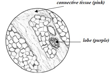
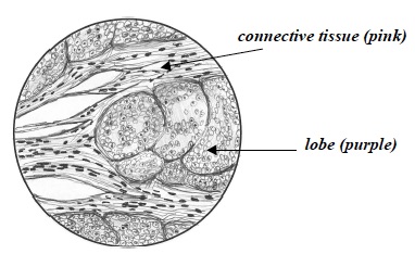
- Review the normal breast tissue slide. Place the slide on the microscope stage. Focus first on the lowest power, and then focus on each of the higher power objectives up to 40X. Do not use the 100X objective.
- Record: Write or draw your observations below when examining a field containing connective tissue and lobe tissue (stained pink and purple respectively) using the 10X objective:
- Place the breast cancer slide on the microscope stage. Focus first on the lowest power, and then focus on each of the higher power objectives up to 40X. Do not use the 100X objective.
- Record: Look at the different cell types and the organization of the cells by moving the microscope stage as needed. Draw a picture of the breast tissue as seen when looking at a field containing connective and lobe tissue with the 10X objective.
- Can you see the lobes and ducts within this breast cancer tissue section? You probably will not be able to see any lobes or ducts in this section. If there are some present they may be filled with cancerous cells or greatly reduced in size.
- Look at the different cell types and the organization of the cells by moving the microscope stage as needed. You may need to switch between the normal and cancer slides multiple times to compare the two tissues and answer the following questions.
- What are the differences in cellular organization in the cancer and normal breast tissues you examined? Are the cells organized or disorganized? In the breast cancer section, you can no longer see the duct and lobe structures seen in normal tissue. The cells are simply disorganized in the cancer slide vs. organized in the normal breast slide.
- How do you think these differences in cellular organization you described will affect the function of the breast? The breast functions to store and transport milk. The lobes and ducts are essential for these functions. The breast will not function normally with the cancer present.
CLEAN UP
Let students know your expectations for clean up. Ask them to clean up.