Teacher Portal:
Cellular Organization
Investigation 3 – Concept Day
ZERO-IN

Italicized font represents information to be shared orally or physically completed with the students at this time.
The non-italicized font represents additional information included to support the teacher’s understanding of the content being introduced within the CELL.
ASK WHY
Remind students that all known living things are made of cells. Studying cells helps us understand how living things function.
BRANCH OUT
Explain to students that stem cell scientists primarily study how stem cells can transform into the various tissues of the human body. Understanding this information can shed light on how to treat cell division ailments, such as cancer and birth defects. Additionally, stem cell scientists look at ways to manipulate this genetic information to grow cells into usable organs and tissues.
PRINT IT
Use your browser to download a printable PDF as a help during the slide presentation and to make additional notes. In your browser, go to File > Print and then choose to save as PDF.
NAVIGATE IT
Once the slide presentation is launched
- use your left and right arrows to advance or go back in the slide presentation, and
- hover your mouse over the left edge of the presentation to get a view of the thumbnails for all the slides so that you can quickly move anywhere in the presentation.
- Click HERE to launch the slide presentation for the CELL.
SHARE IT
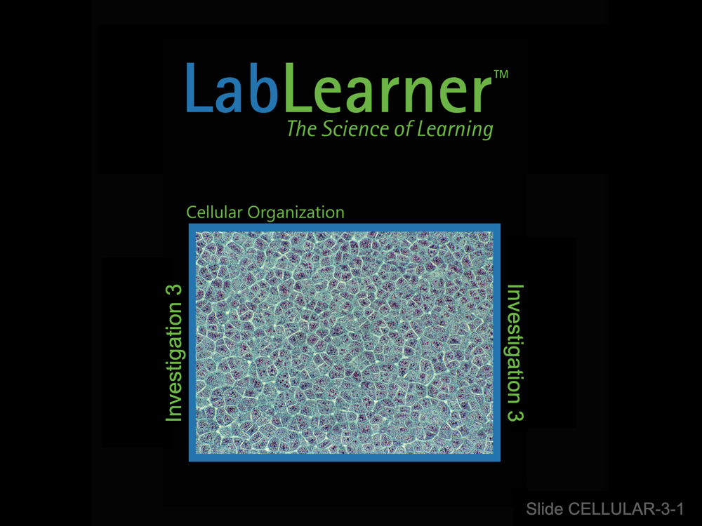
SLIDE CELLULAR-3-1
Note: In Lab, students will compare plant and animal cells using the light microscope. Students will be able to see the primary differences between plant and animal cells first hand. They will conclude that there are many similarities between the two types of cells but that plant cells are unique in having chloroplasts and a cell wall. They may also observe that plant cells have a large, dominant vacuole in their cytoplasm whereas animal cells do not.
In order to see greater detail than that described above, it takes significantly more magnification and resolution. This is achieved in practice using the electron microscope. Only with this instrument can one clearly differentiate between different organelles within the cytoplasm of cells. Therefore, most of this presentation is devoted to showing students high magnification photographs of plant and animal cells.
We also present an “idealized” model of both an animal and a plant cell. However, we attempt to show that such models are not derived from images like the students will have observed through their microscopes, but through the use of electron microscopy. Once the plant and animal cell models are presented, their differences and similarities are depicted and a Venn diagram is presented. The final slide addresses the cellular functions of some of the organelles shown in models.
________________________________________
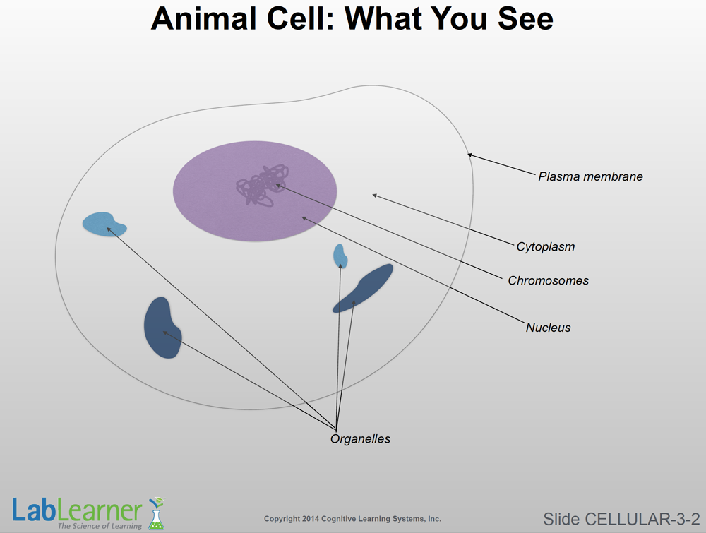
SLIDE CELLULAR-3-2
- Animal Cell: What You See. This simple slide shows the amount of detail that students are likely to see in animal cell preparations through their light microscopes. While chromosomes are indicated on the illustration, these structures are only seen clearly in light microscopy in certain preparations of actively dividing cells.
Note: Students will become much more familiar with the nucleus and chromosomes in the upcoming CELL, Cell Cycle, and Cancer.
- As mentioned above, it is difficult or impossible to differentiate between the various cell organelles with the light microscope.
________________________________________
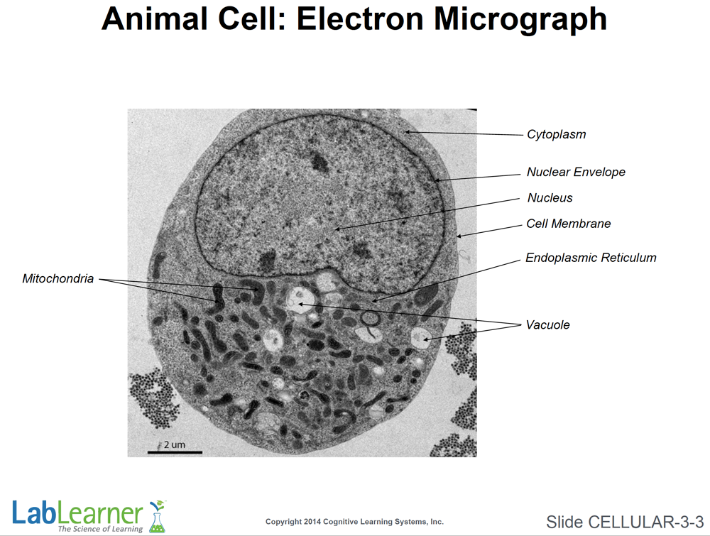
SLIDE CELLULAR-3-3
- Animal Cell: Electron Micrograph. This slide shows a Transmission Electron Microscope image of an animal cell with some of its organelles labeled. There are many mitochondria in this cell. By comparing many such images, scientists have been able to construct models of animal cells like the one shown in the next slide.
________________________________________
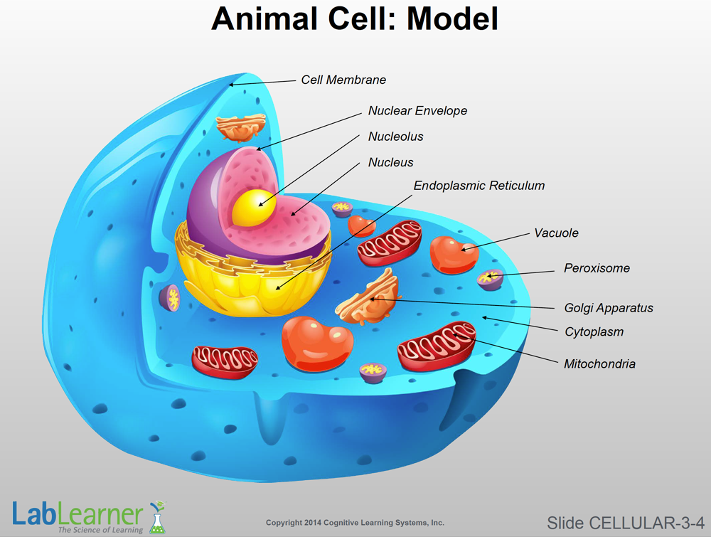
SLIDE CELLULAR-3-4
- Animal Cell: Model. This slide shows an idealized model of an animal cell. It is not meant to represent any particular type of animal cell and all animal cells are somewhat different from each other. It therefore simply depicts the common sub-cellular structures found in greater or lesser abundance in all animal cells.
Note: The final slide of this presentation includes a brief description of the functions of the various organelles shown in this model. It is also presented below for reference:
_____________________________________
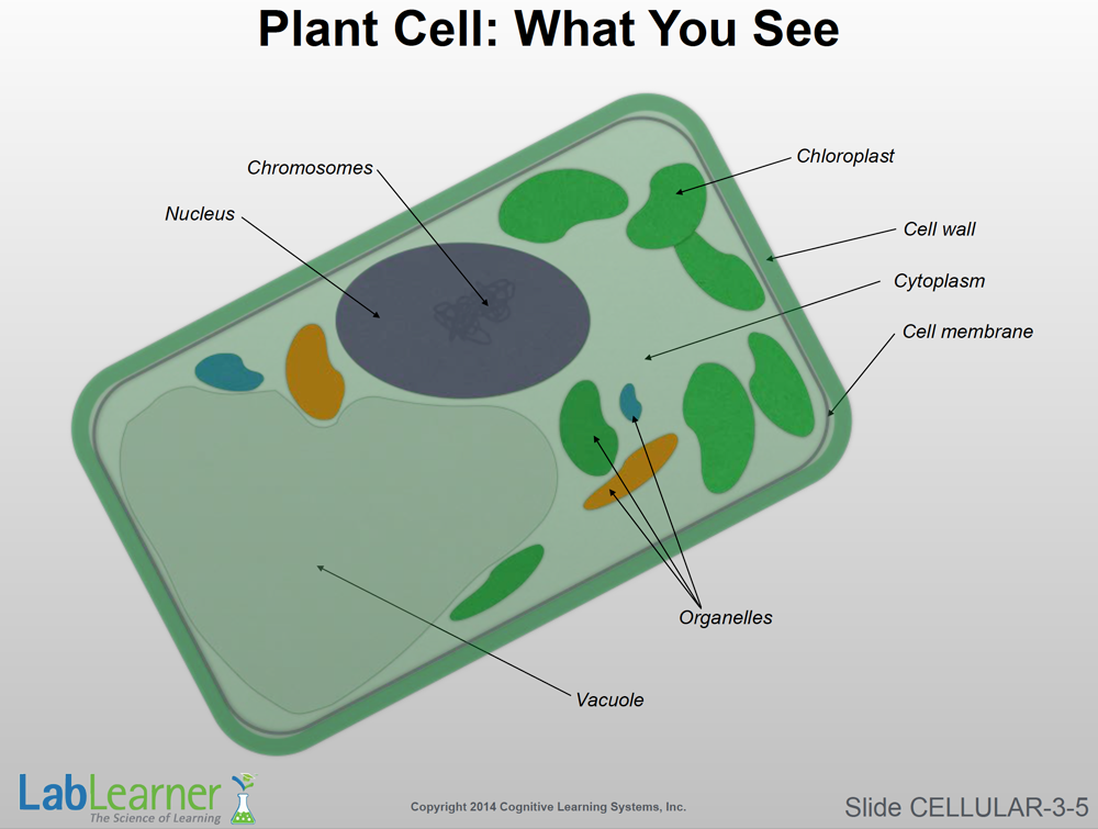
SLIDE CELLULAR-3-5
- Plant Cell: What You See. Turning now from animal to plant cells, this slide represents the structures that students are likely to see through their light microscopes.
Note: As in the case with the animal cell, students will not be able to differentiate between the various organelles with the notable exception of the chloroplasts.
- Chloroplasts are the cellular site of photosynthesis and usually show up well in the light microscope. The cell wall and large water-filled vacuole should also be seen by students in the lab.
_____________________________________
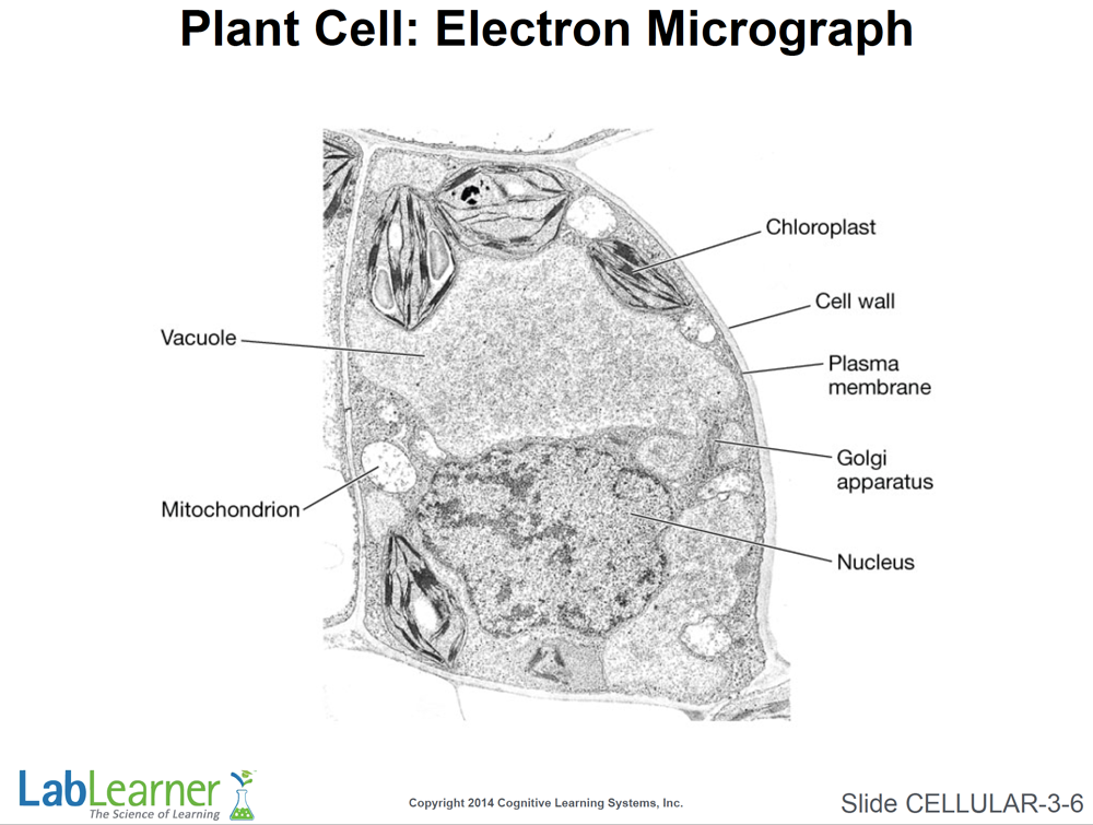
SLIDE CELLULAR-3-6
- Plant Cell: Electron Micrograph. This is a Transmission Electron Microscope image of a plant cell.
-
Note: The Golgi apparatus and mitochondrion (singular for mitochondria) will not be seen by students in the lab through the light microscope.
Note: There are other sub-cellular structures not labeled on this micrograph. Plant scientists have constructed models by examining many micrographs like the one shown here.
_____________________________________
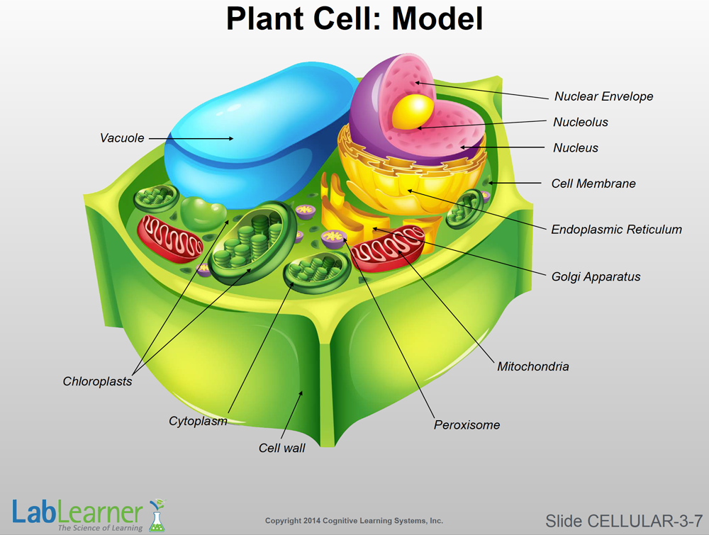
SLIDE CELLULAR-3-7
- Plant Cell: Model. This slide shows an idealized model of a plant cell. It is not meant to represent any particular type of plant cell and all plant cells are somewhat different from each other. It therefore simply depicts the common sub-cellular structures found in greater or lesser abundance in all plant cells.
_____________________________________
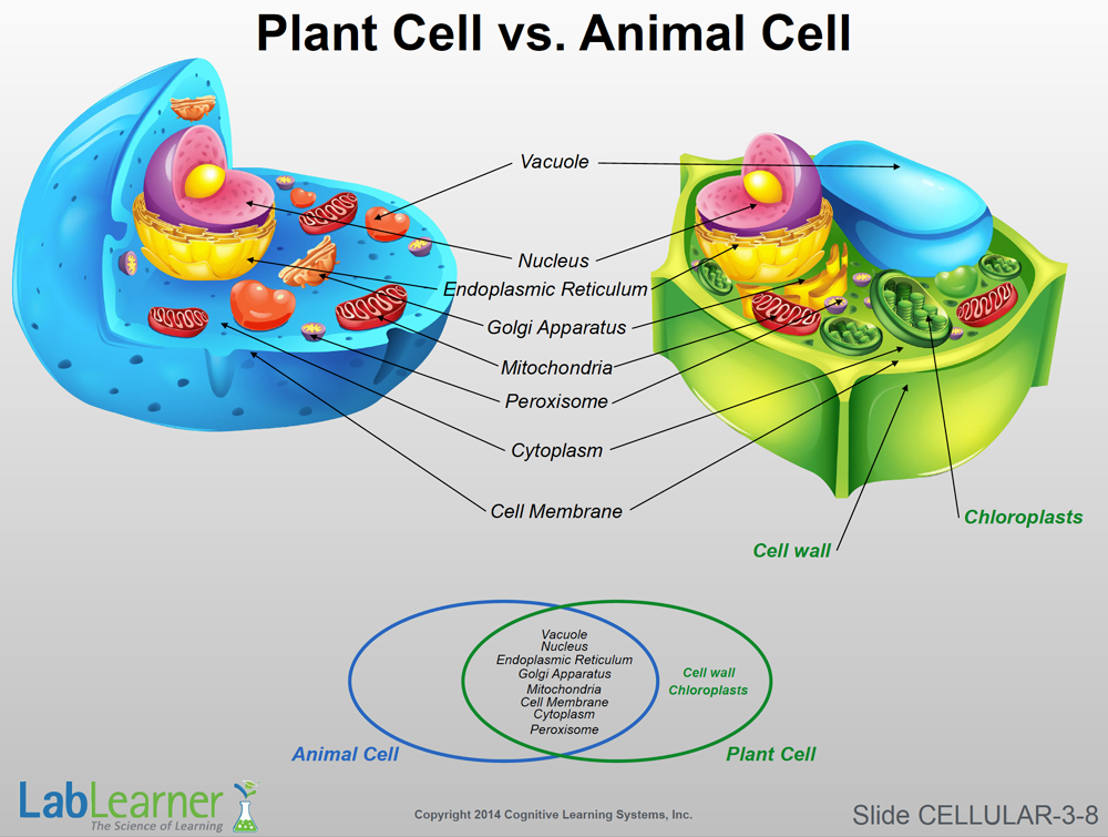
SLIDE CELLULAR-3-8
- This slide simply shows the animal and plant cells together. Notice that each cellular structure, with the exception of the cell wall and chloroplasts, are present in both plant and animal cells.
- At the bottom of the slide, a Venn diagram is presented that once more highlights the similarities and differences between plant and animal cells.
_____________________________________
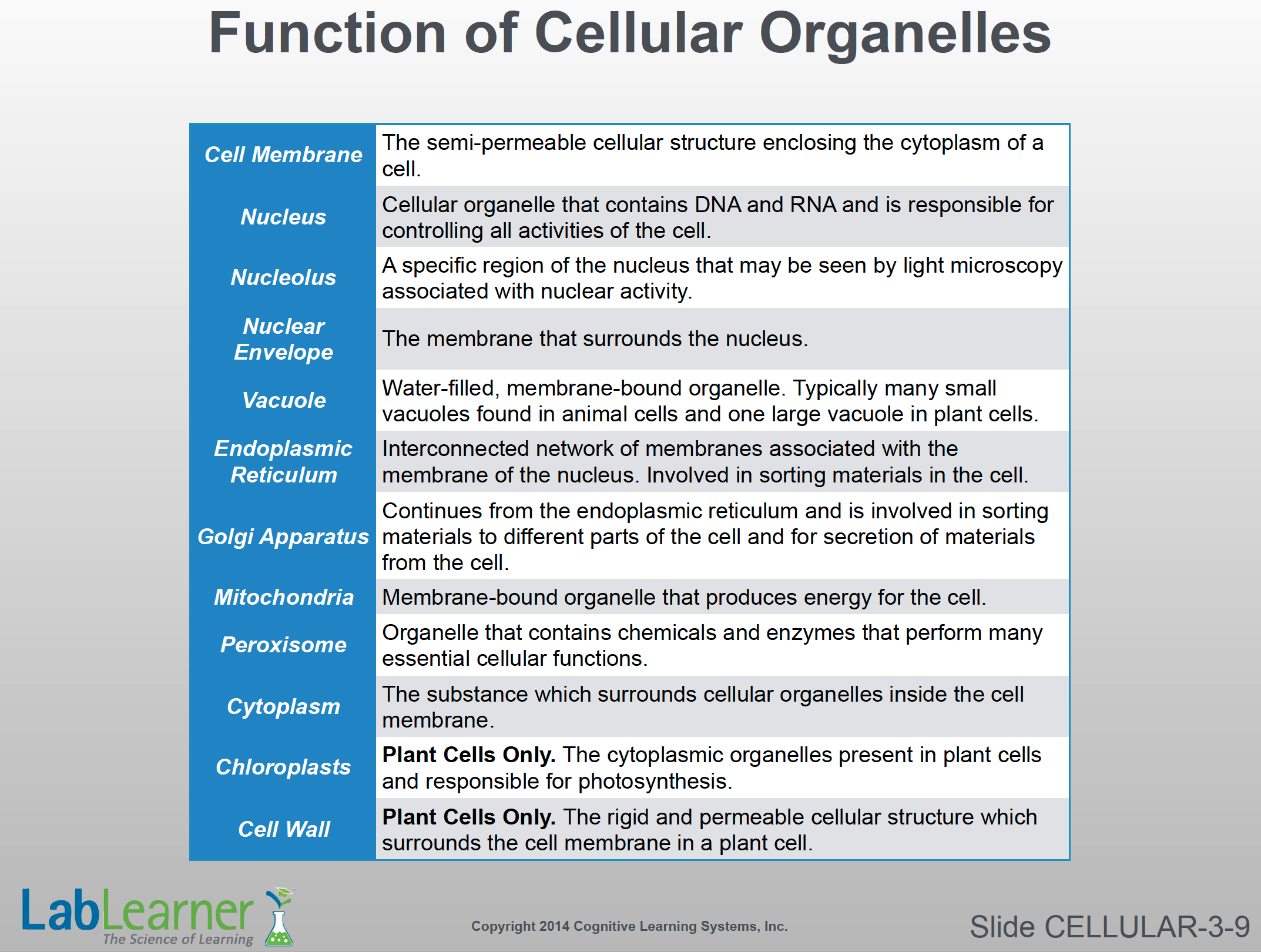
SLIDE CELLULAR-3-9
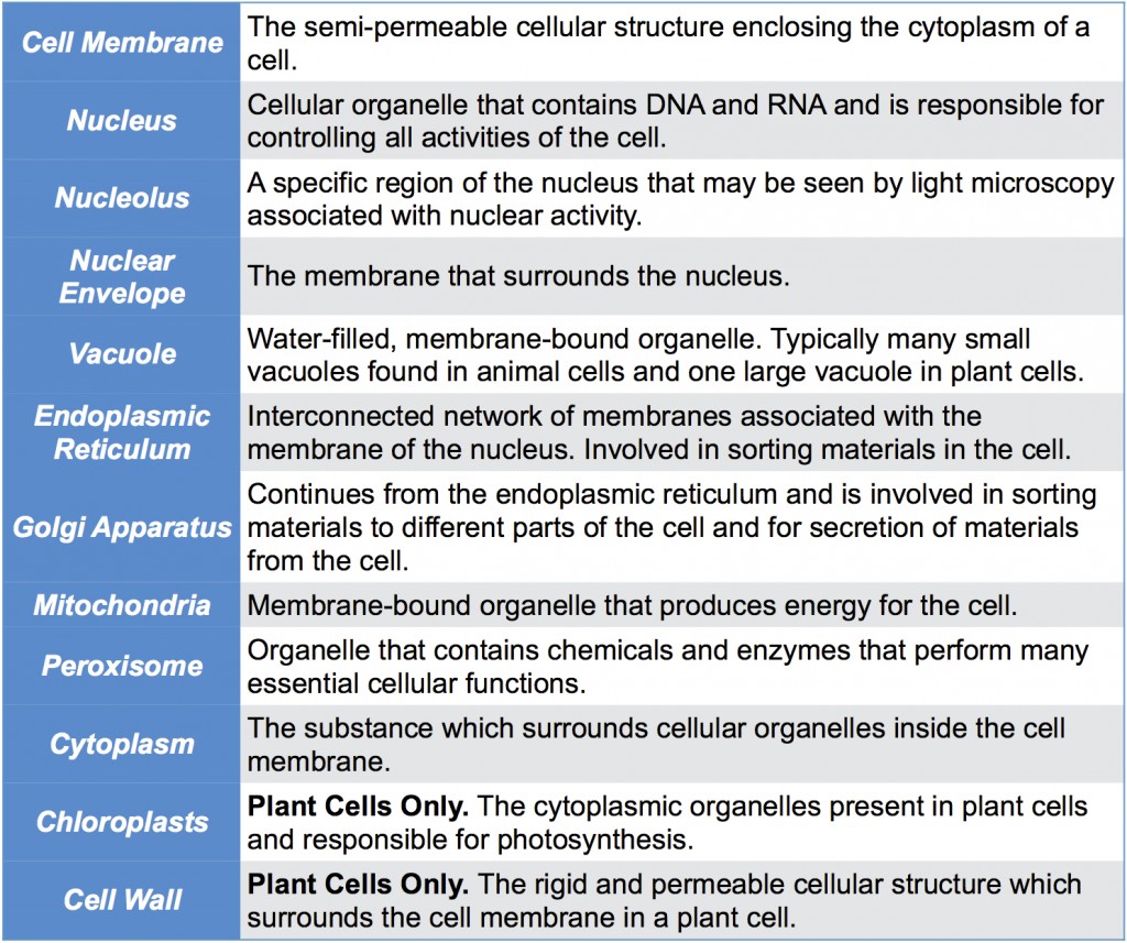
Note: This slide simply lists the basic function of each of the cell organelles labeled or discussed in this presentation.