Teacher Portal:
Microscopes and Magnification
Investigation 4 – PostLab
 ASK WHY
Microscopes have made a tremendous contribution to science since their use began in the sixteenth century (the 1500s).
Microscopes are one of the most important scientific instruments developed. In fact, in the medical field, microscopes are largely responsible for making modern medicine “modern”!
BRANCH OUT
Microscopists today work in many different fields including field and laboratory life sciences, chemistry, materials science, and nearly every branch of biomedical research and medicine.
PRINT IT
Use your browser to download a printable PDF as help during the slide presentation and to make additional notes. In your browser, go to File > Print and then choose to save as PDF.
NAVIGATE IT
Once the slide presentation is launched
- use your left and right arrows to advance or go back in the slide presentation, and
- hover your mouse over the left edge of the presentation to get a view of the thumbnails for all the slides so that you can quickly move anywhere in the presentation.
- Click HERE to launch the slide presentation for the CELL.
SHARE IT
SLIDE M&M4-post-1
Begin the PostLab Analysis by asking students to recall the experiments they conducted. The questions on the following slide will help prompt student discussion.
______________________________________________
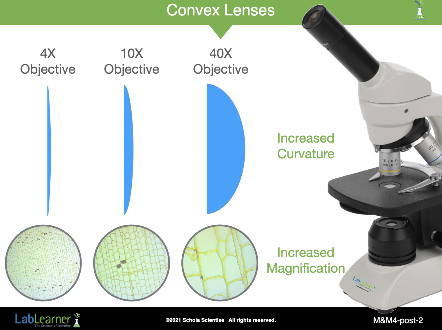
SLIDE M&M4-post-2
Ask the following questions comparing the hand lens and the compound microscope.
Why are the hand lens and the compound microscope able to magnify an object’s image? The hand lens and the microscope both have lenses. Since surfaces of the lenses are curved, light is refracted when it passes through the lenses.
What type of lens do the hand lens and the microscopes objectives contain? Each lens is a convex lens.
How do you know that they are convex and not concave? The lenses are convex since the images produced by the hand lens and by the microscope are magnified. A concave lens would have reduced the size of the image.
How does a convex lens magnify an object’s image? The light reflected off of the object is refracted so that the image of the object is magnified, increasing the apparent size of the object.
What is the difference between the model of the low power objective using the hand lens and the compound microscope’s low power objective? The hand lens model of a low power objective did not magnify the image as much as the low power objective of the microscope.
- Inform students that there are two reasons the power of magnification is different for the hand lens and the microscope. The lens in the microscope objective is more curved than the curvature of the lens in the hand lens. A greater curvature results in greater refraction of light resulting in a larger image. Also, the total magnification of the microscope is the combination of the lenses in the objective and in the eyepiece.
______________________________________________
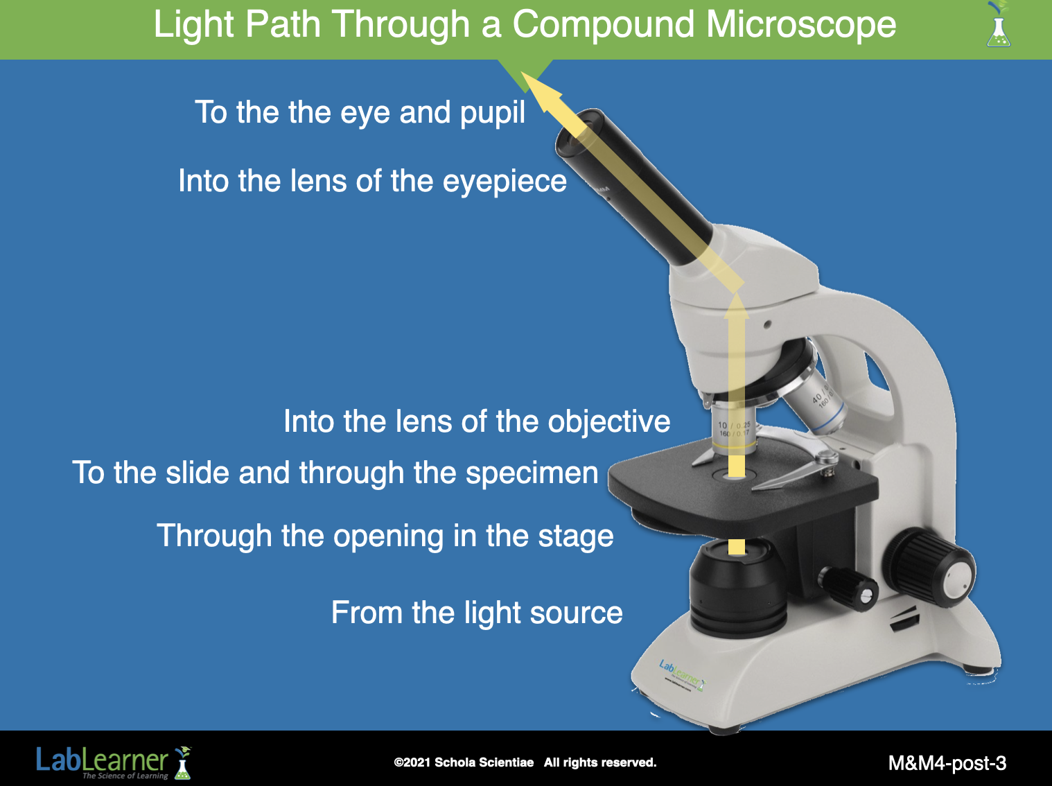
SLIDE M&M4-post-3
This slide provides an opportunity to trace the path of light through a compound microscope. Light travels:
1. From the light source.
2. Through the opening on the stage.
3. To the slide and through the specimen.
4. Into the lens of the objective.
5. Into the lens of the eyepiece.
6. Where the eye and its pupil allow light to enter.
______________________________________________
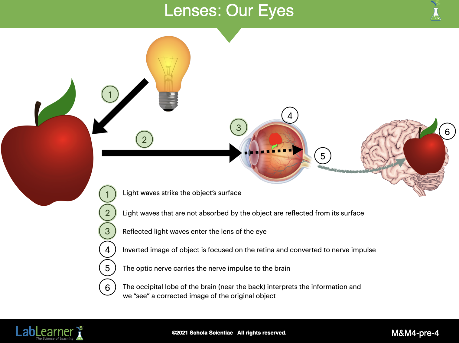
SLIDE M&M4-post-4
Ask students to recall that the light enters the eye and triggers a nerve signal at the back of the eye. The nerve signal is carried to the brain by the optic nerve. The brain then identifies the image.
- Explain to students that light reflects off of an object and then passes through the outer layer of the eye, called the cornea.
- The light then enters the eye through an opening called the pupil. The size of the pupil is controlled by the iris, which is the colored part of the eye.
______________________________________________
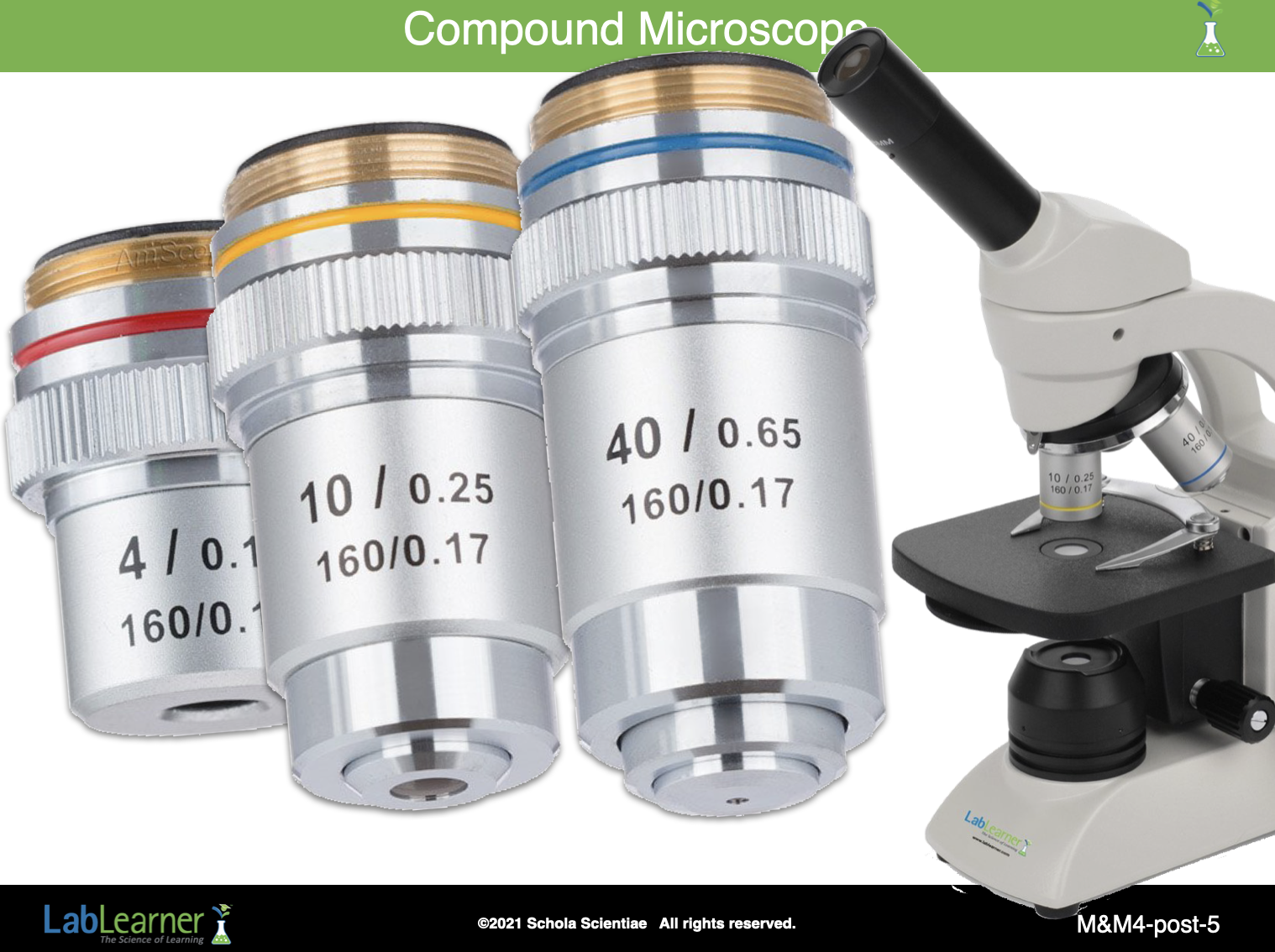
SLIDE M&M4-post-5
Ask the following questions about how the compound microscope functions:
Which objective of the compound microscope allows you to see less detail, but a larger field of view? The lowest objective allows you to see less detail but an increased field of view.
Which objective of the compound microscope allows you to see more detail, but a smaller field of view? The highest objective allows you to see more detail but a decreased field of view.
You should always begin viewing a specimen with which objective of the compound microscope? Why does this procedure protect the slide? The lowest power objective (4x) should be used first. If the medium (10x) or high (40x) power objectives are used first, there is a chance of breaking the slide when focusing on the specimen since these objectives are longer.
What is another reason to begin viewing a specimen using the lowest power objective? Student answers will vary. The greater field of view of the lowest power objective allows the viewer to center the objective on the specimen. If the specimen is viewed first with the highest power objective, its small field of view could result in missing the specimen.
______________________________________________
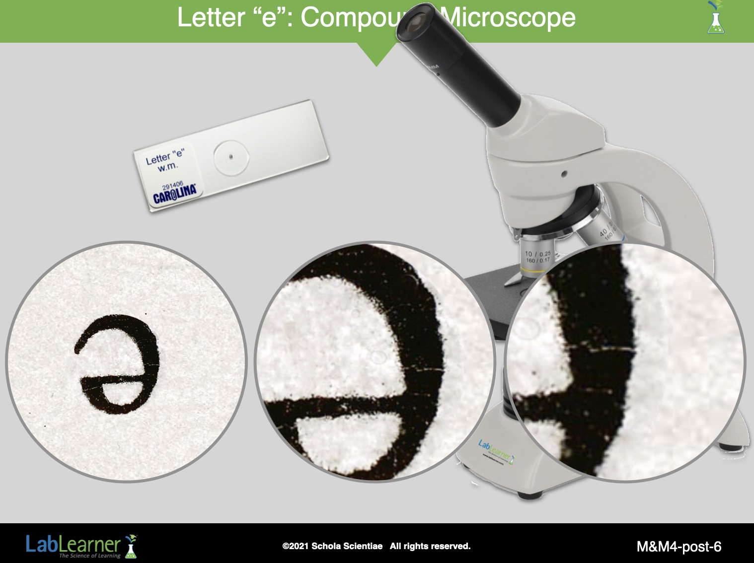
SLIDE M&M4-post-6
Begin a discussion of students’ observations of the “letter e” slide. Direct students to Problem 5 in their Student Data Record.
Ask students to describe the image of the letter “letter e” when viewed using each of the three objectives:
Describe the image of the “letter e” slide when viewed using the low power (4x) objective. Students will describe the image as being larger or more detailed than the slide itself.
Describe the field of view. The field of view was smaller.
Describe the image of the “letter e” slide when viewed using the medium power (10x) objective. Students will describe the image as being larger or more detailed than when viewed using the 4x objective.
Could you view all of the image? No, only a portion of the image could be viewed.
Describe the field of view when using the medium power objective as compared to the low power objective. The field of view was smaller than when using the 4x objective.
Describe the image of the “letter e” slide when viewed using the high power (40x) objective. Students will describe the image as being larger or more detailed than when viewed using the 10x objective.
Could you view all of the image? No, only a small portion of the image could be viewed.
Describe the field of view when using the high power objective as compared to the medium power objective. The field of view was smaller than when using the 4x objective.
______________________________________________
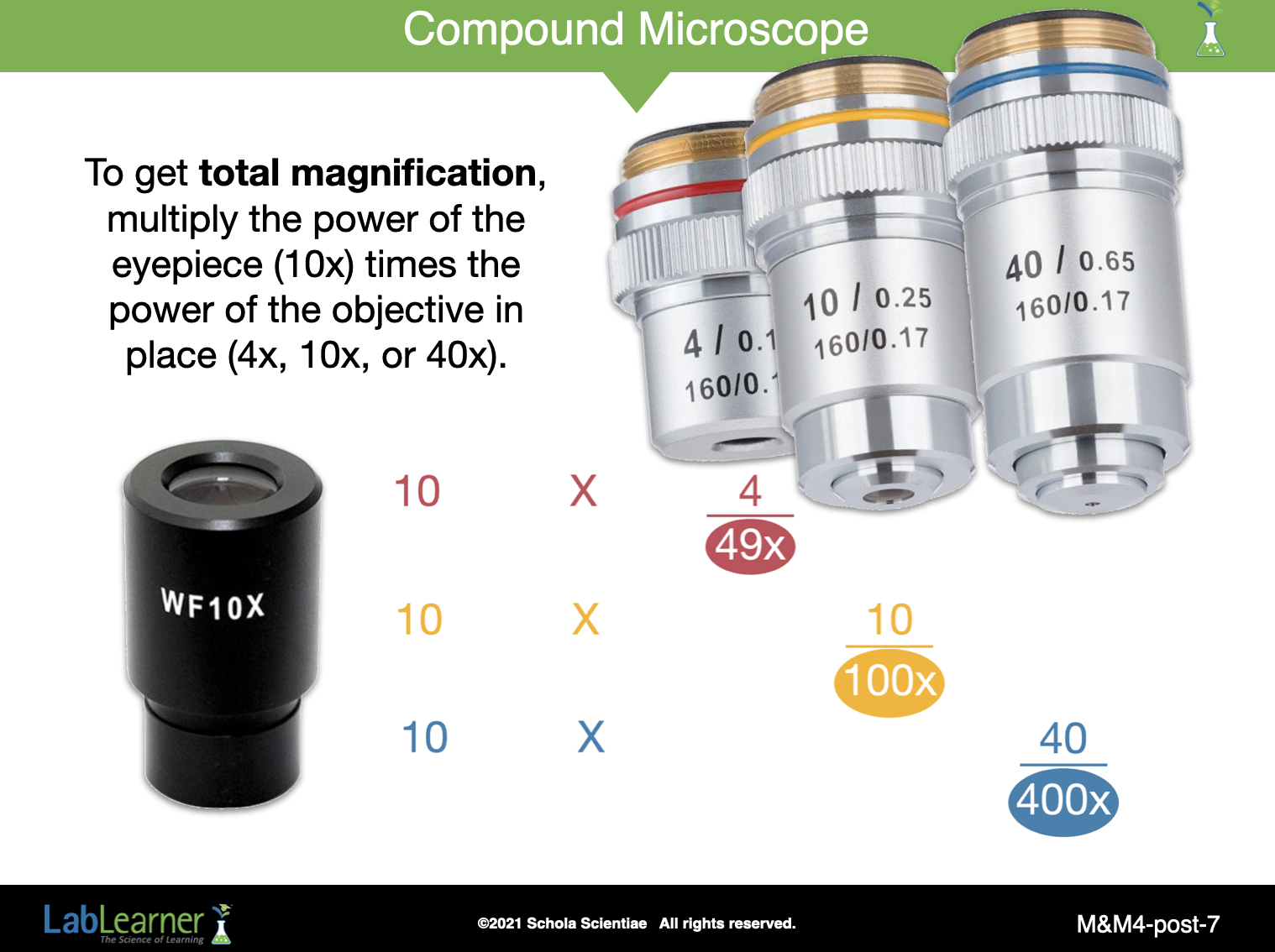
SLIDE M&M4-post-7
Review how to calculate the total magnification of each objective of the compound microscope. This slide has been seen before and should facilitate discussion of how to calculate the power of magnification.
Remember, the total magnification is calculated by multiplying the magnification of the objective by the magnification of the eyepiece.
______________________________________________
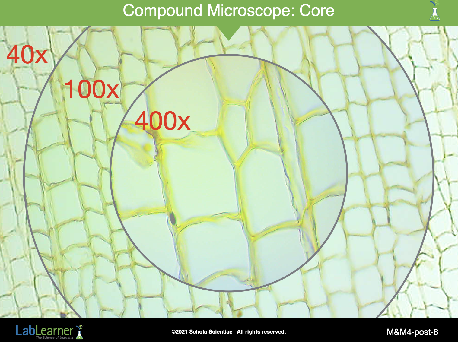
SLIDE M&M4-post-8
Begin a discussion of students’ observations of the cork slide. Direct students to Problem 6 in their Student Data Record. This slide shows three different images of the prepared cork slide with the total magnification indicated for each image.
Ask students to describe the image of the cork when viewed using each of the three objectives.
Describe the image of the cork slide when viewed using the low power (4x) objective. Students will describe the image as being larger or more detailed than the slide itself.
Describe the field of view. The field of view was smaller.
Describe the image of the cork slide when viewed using the medium power (10x) objective. Students will describe the image as being larger or more detailed than when viewed using the 4x objective.
Could you view all of the image? No, only a portion of the image could be viewed.
Describe the field of view when using the medium power objective as compared to the low power objective. The field of view was smaller than when using the 4x objective.
Describe the image of the cork slide when viewed using the high power (40x) objective. Students will describe the image as being larger or more detailed than when viewed using the 10x objective.
Could you view all of the image? No, only a smaller portion of the image could be viewed.
Describe the field of view when using the high power objective as compared to the medium power objective. The field of view was smaller than when using the 10x objective.
______________________________________________
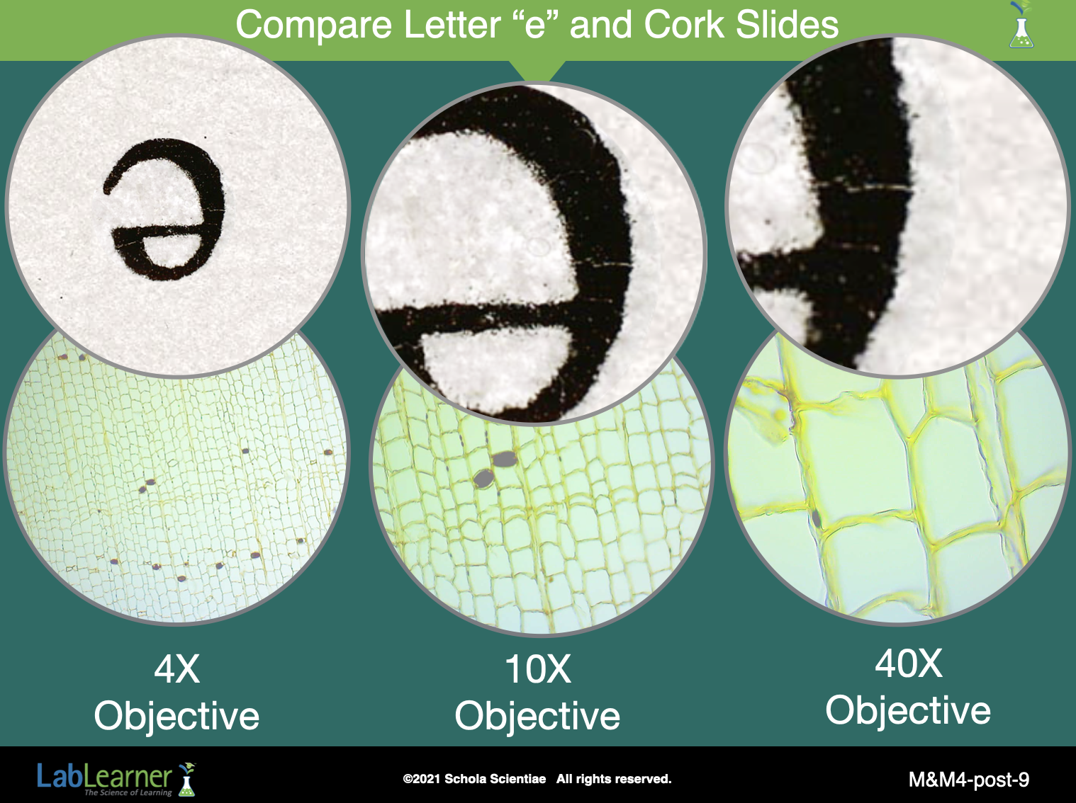
SLIDE M&M4-post-9
Ask students to summarize their observations of the two slides using each of the three objectives. Students should refer to their answers to Problems 7 and 8 in their Student Data Records:
- Low power (40x): Student answers will vary. Most of the specimen is visible (a large field of view). Not many details are visible.
- Medium power (100x): Student answers will vary Less of the specimen is visible (an intermediate field of view), but more details are visible.
- High power (400x): Student answers will vary. Very little of the specimen is visible (a small field of view), but more details and textures are visible.
Does the compound microscope or the model of the low-power objective lens produce more or less light refraction? Why? The compound microscope produces greater light refraction because it is capable of higher total magnification (400x) than the model of the low power objective (1.17x, see the following slide). The greater the magnification, the more that light is refracted.
______________________________________________
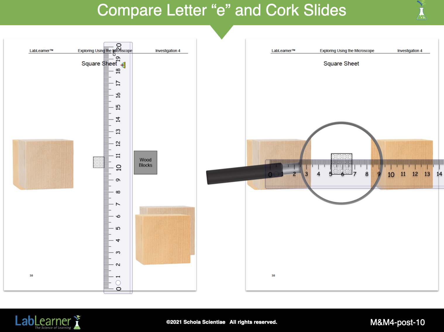
SLIDE M&M4-post-10
Discuss how to calculate the power of magnification of the hand lens.
Ask students to turn to the Table in their Student Data Record and review the data they recorded in the Lab.
What was the width of the square when viewed by the naked eye? Approximately 1.2 centimeters.
Did the square appear larger or smaller when using the hand lens? Larger
What was the width of the square’s image when viewed using the hand lens? Approximately 1.4 centimeters.
How can you calculate the power of magnification of the hand lens? Student answers will vary.
______________________________________________
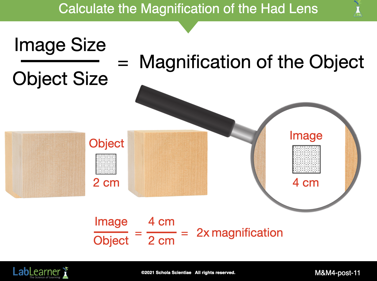
SLIDE M&M4-post-11
Direct students to Problem 4 in their Student Data Record.
• A square has a width of 2 centimeters. If a lens increases the width of the image to 4 centimeters, what is the power of magnification of that lens? 2x magnification
Finally, using actual classroom data shown on the previous slide ( object = 1.2 cm, image = 1.4 cm), Calculate the magnification of the hand lens.

______________________________________________