Teacher Portal:
Microscopes and Magnification
Performance Assessment – Lab
 BE PREPARED
Materials
Class materials:
-
- Masking tape
Group materials
-
- 1 compound microscope
- 1 pinus stem slide
- 1 mammalian intestine slide (colon)
- 1 human blood smear slide
Individual materials
-
- Student Data Record
TEACHER PREPARATION
- Cover the labels on each slide with a small piece of masking tape
- Label the pinus stem slides “1”.
- Label the mammalian intestine slides “2”.
- Label the human blood smear slides “3”.
- Divide the class into five groups of students
INSTRUCTION
1. Instruct each student group to obtain the following materials from the distribution point: one (1) compound microscope, one (1) slide 1, one (1) slide 2, and one (1) slide 3.
PROCEDURE
 1. Begin the Performance Assessment by reading the opening paragraph from the Scientist Data Record aloud. Briefly review the contents of the paragraph and outline the goals of this project to the class. Read aloud the goals from the list on the Scientist Data Record.
2. Inform students that they will make observations of the three specimen slides using the compound microscope. Only one of the slides is blood.
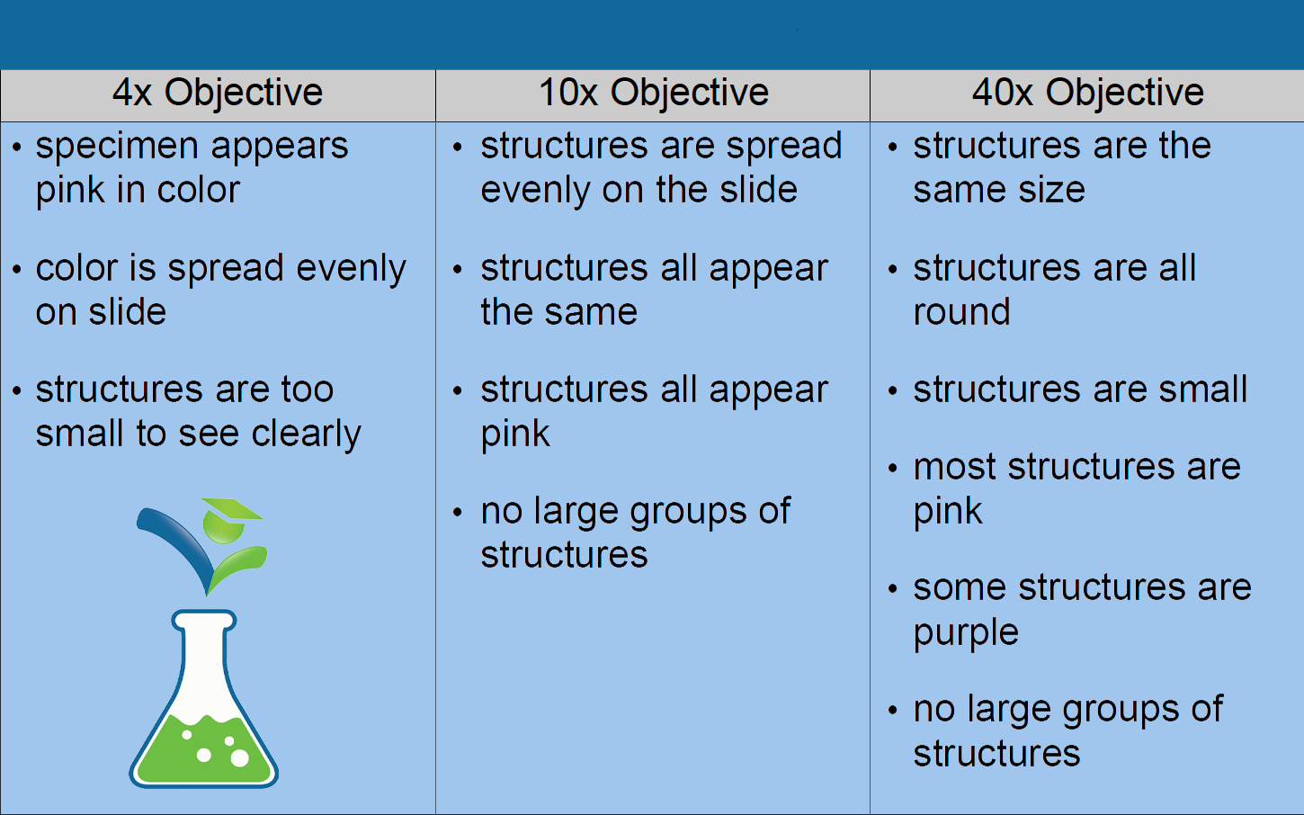
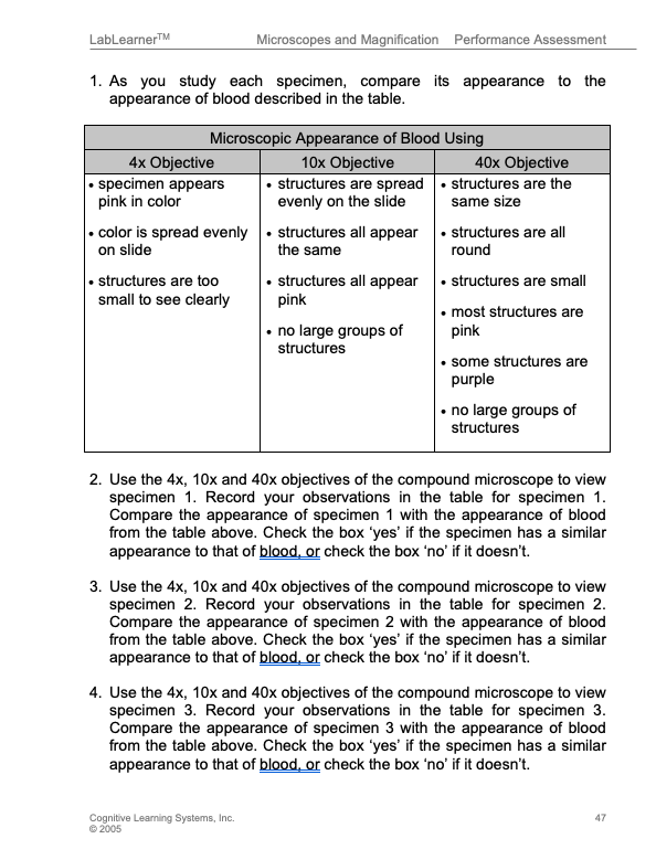
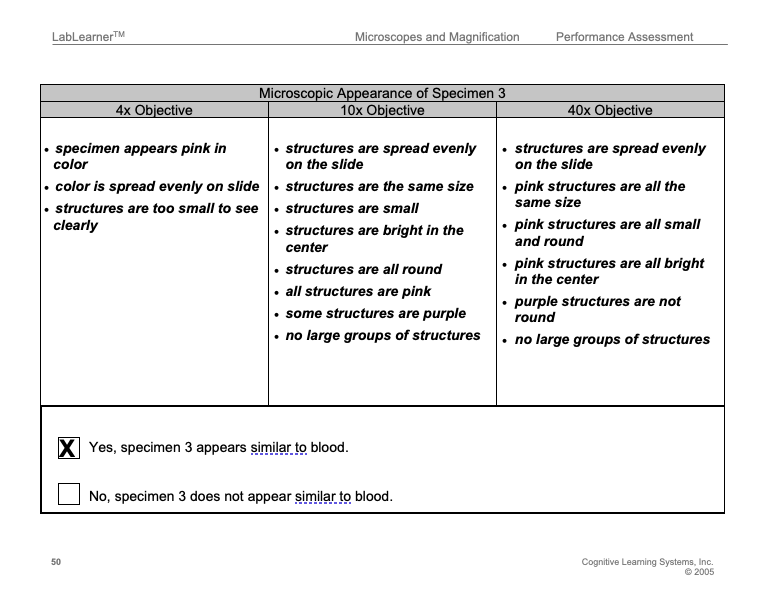
3. Inform students that to guide them in their observations of the specimens, they should use the table in Problem 1 of their Student Data Record. The table includes descriptions of the microscopic appearance of blood when using the 4x, 10x, and 40x objectives.
4. Before students begin the Performance Assessment, walk through each step and encourage students to verbalize their plans for how to solve the problem. Make certain that students view each specimen using each of the three objectives of the compound microscope.
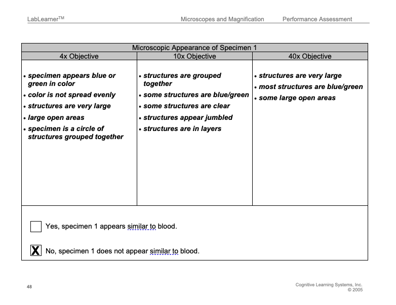
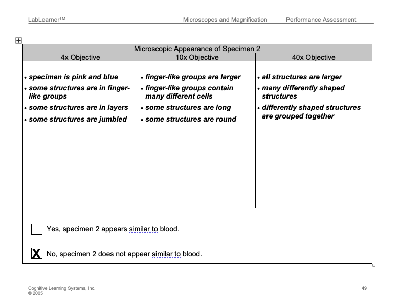
5. Students should record their observations of the three specimens using each objective in the tables in their Student Data Records.
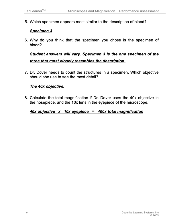
6. Based on their recorded observations, students should record in problem 5 in their Scientist Data Record which specimen they believe is the specimen of blood.
 NOTES: Students will likely not have prior knowledge of cells and do not need this knowledge in order to complete the Performance Assessment successfully. Students simply compare the structures visible in each specimen. The following description is provided for teacher background:
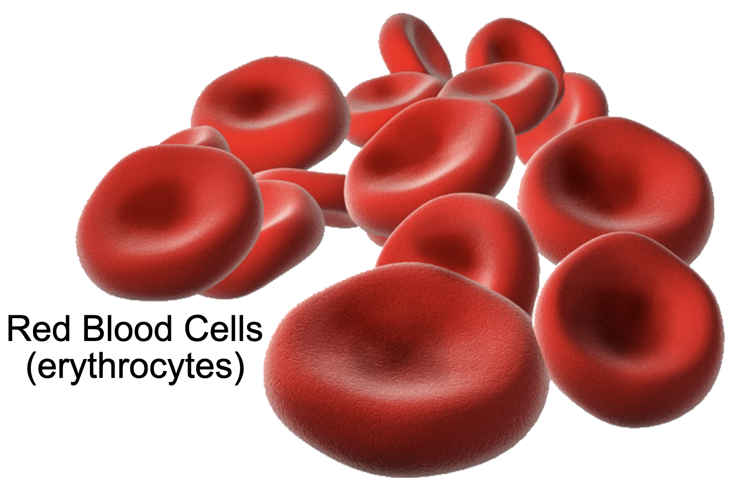
Blood mainly contains red blood cells (erythrocytes) which contain the hemoglobin necessary for transporting oxygen. These cells are small, pink, round cells. Blood also contains a smaller number of white blood cells (leukocytes) that help the body fight infections. The white blood cells appear purple because of the staining procedure.
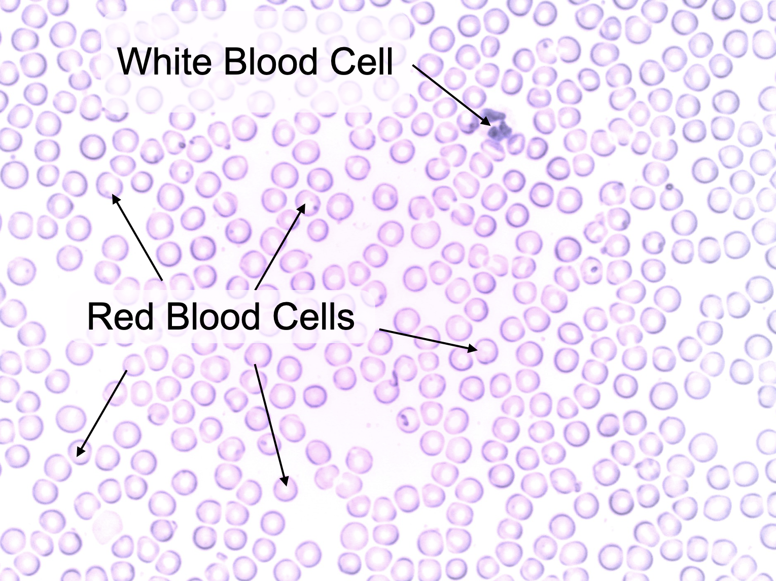 The blood smear is stained with two different dyes: a pink dye that stains the red blood cells and a purple dye that stains the nucleus of every white blood cell. The red blood cells are not stained with the purple dye because they do not contain a nucleus.
Unlike specimens 1 and 2, blood does not contain groups of cells that form definite structures. The pinus stem and the mammal intestine both have definite structures made up of groups of different types of cells. These structures are cells of different types organized into tissue. Students will learn much more about tissues in Microscopic Explorations and the middle school CELL, Cellular Organization.
7. Students should then answer Problems 6 – 8 in their Student Data Record.
KEYS: PERFORMANCE ASSESSMENT
CLEAN UP
Let students know your expectations and instruct them to clean up their lab bench after the Performance Assessment.