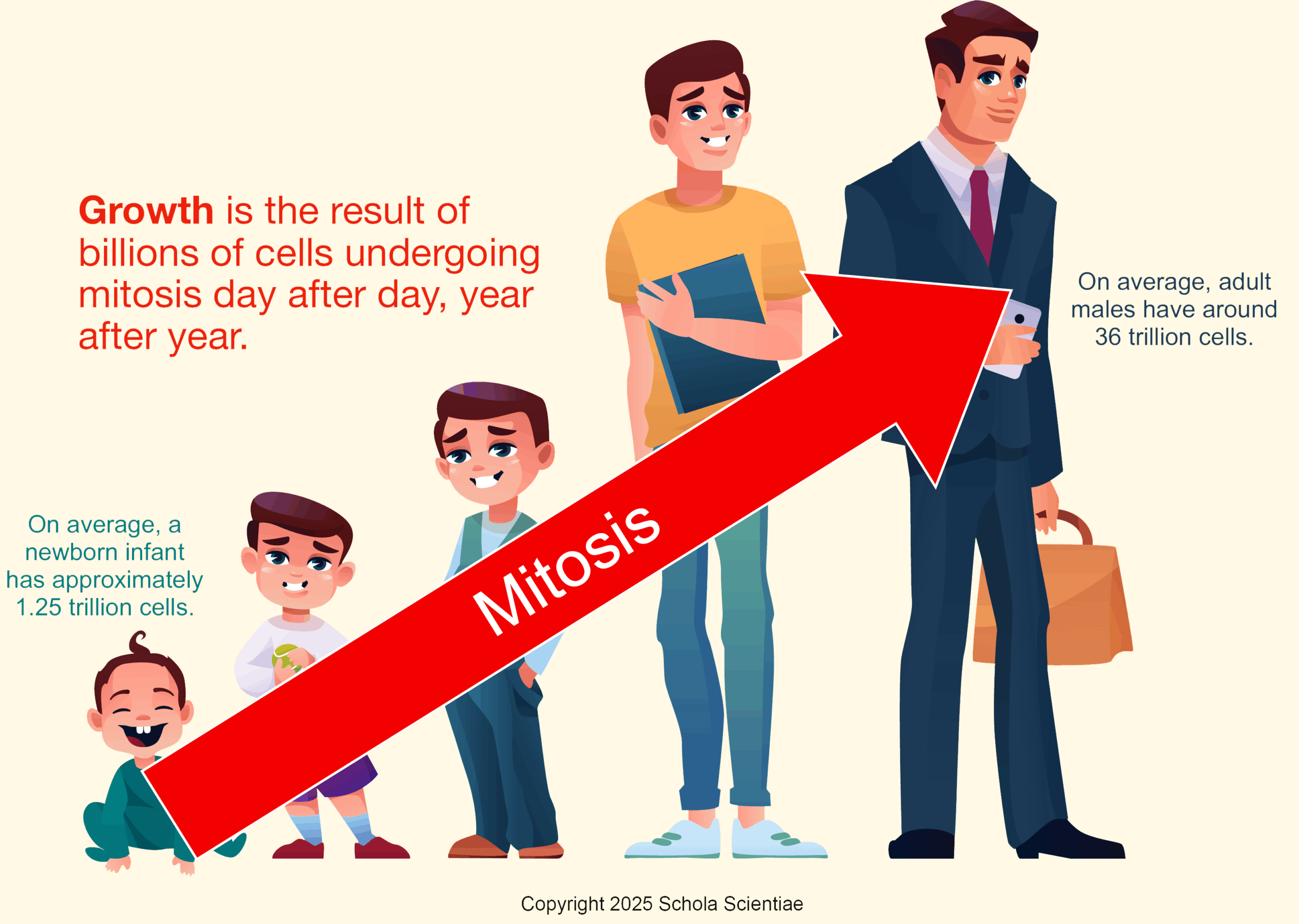
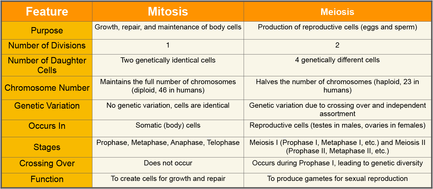
Mitosis and Growth

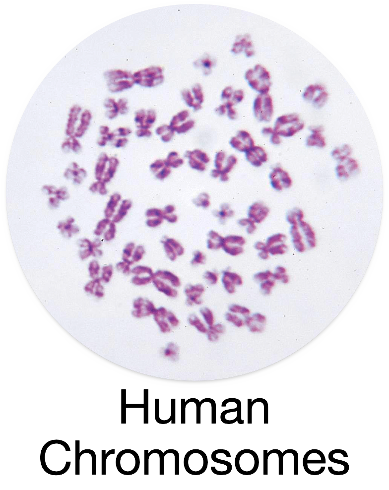
Human Chromosomes
Think of chromosomes like tiny instruction books that tell our bodies how to grow, develop, and work. We have 46 chromosomes in total, organized into 23 pairs. One pair of these chromosomes is called the sex chromosomes, and they determine whether someone is male or female.
 Male and Female Chromosomes
- Females have two X chromosomes, written as XX.
- Males have one X chromosome and one Y chromosome, written as XY.
How Do They Work?
When a baby is made, it gets one chromosome from each parent to form the pair. The mother always gives an X chromosome because that’s all she has (XX). The father can give either an X or a Y chromosome.
- If the father gives an X chromosome, the baby will be female (XX).
- If the father gives a Y chromosome, the baby will be male (XY).
So, the father’s chromosome is the one that decides if the baby will be a boy or a girl.
In short:
- Females have XX chromosomes.
- Males have XY chromosomes.
- It’s the father’s chromosomes that determine the baby’s sex.
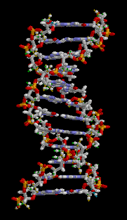
- Finally, each chromosome contains long strands of the deoxyribonucleic acid (DNA) molecule. The DNA strand is organized into sequences called genes, each of which codes for specific proteins that proform necessary biochemical functions in our cells.
Check Your Understanding
 Q: How many chromosomes do humans have?
A: Humans have 46 chromosomes, organized into 23 pairs.
- Q: What determines if a baby will be male or female?
A: The father’s sperm determines sex: an X chromosome means female (XX), and a Y means male (XY).
- Q: What is the role of DNA in chromosomes?
A: DNA contains genetic instructions for growth, development, and cell function.
- Q: What is a micrograph?
A: It’s an image taken through a microscope, often used to study chromosomes.
- Q: How do chromosomes pass from parents to children?
A: Each parent contributes one chromosome per pair during fertilization.
Check Your Understanding
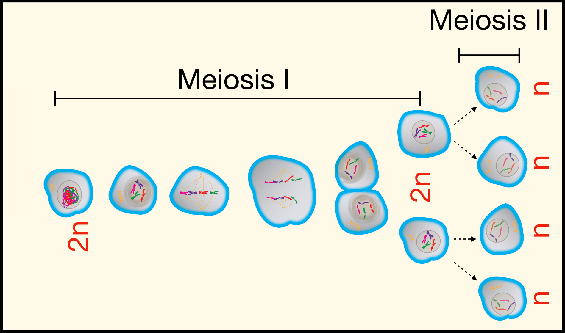
1. Q: How does meiosis differ from mitosis?
A: Meiosis creates four genetically different cells with half the usual number of chromosomes for reproduction.
2. Q: Why do human cells have 46 chromosomes?
A: Each parent contributes 23 chromosomes during fertilization, totaling 46.
3. Q: How does genetic diversity occur in meiosis?
A: It happens through chromosome shuffling and crossing over during meiosis I.
Menstrual Cycle and Reproduction
To understand fertilization, you need to know about the menstrual cycle. The menstrual cycle happens roughly every month in a female’s body to prepare for a possible pregnancy.
The human menstrual cycle typically begins during puberty, which is when a female’s body starts to develop and change. The usual age for this to start is around 12 years old, but it can happen anytime between 9 and 16 years old.
Once the menstrual cycle starts, it generally continues each month until a person reaches menopause, which usually happens between the ages of 45 and 55, when the menstrual cycle stops permanently.
So, the overall age range for the menstrual cycle is roughly 9 to 55 years old, though it can vary slightly from person to person.
The menstrual cycle usually lasts about 28 days but can be a little shorter or longer. Here’s a simple overview:
Days 1-5: The cycle begins with menstruation when the lining of the uterus (the womb) sheds if no fertilization happened in the previous cycle. This lining exits the body as what we call a “period.”
Days 6-14: The body starts to prepare for the next cycle. A new egg cell begins to grow and mature in the ovary. Meanwhile, the lining of the uterus starts to thicken again, getting ready for a fertilized egg to attach.
Around Day 14 (Ovulation): This is the ovulation phase. The egg leaves the ovary and travels down the fallopian tube, where it might meet a sperm. This is the time when fertilization is most likely to happen because the egg is ready to be fertilized.
Days 15-28: If the egg is not fertilized, it breaks down, and the uterus lining gets ready to shed again, starting a new cycle.
Fertilization can happen if sperm enters the female body during or close to the time of ovulation (around Day 14). Sperm cells swim through the uterus to reach the fallopian tube, where they might meet the egg. When one sperm manages to join with the egg, it fertilizes it, and the egg becomes a zygote, the very first stage of a new human life.
After fertilization, the zygote travels to the uterus and attaches to the thickened lining. This is where it will continue to grow and develop into a baby over the next nine months.
In summary:
- Fertilization can only occur if sperm meets the egg when it’s in the fallopian tube, which happens around Day 14 of the menstrual cycle.
- If fertilization doesn’t happen, the egg will eventually break down, and the cycle starts again.
____________________________________
Fertilization
Fertilization is the process where a sperm cell from the father combines with an egg cell from the mother to form a new, genetically unique individual. Only males can produce sperm cells, and only females can produce egg cells (also called ova). Meiosis is required to form both types of gametes (sex cells).
Both sperm and egg cells are haploid, meaning they each have only half the number of chromosomes needed to make a complete set. A complete set of chromosomes in humans is 46, but each haploid cell has only 23 chromosomes.

The Role of Sperm and Egg Cells
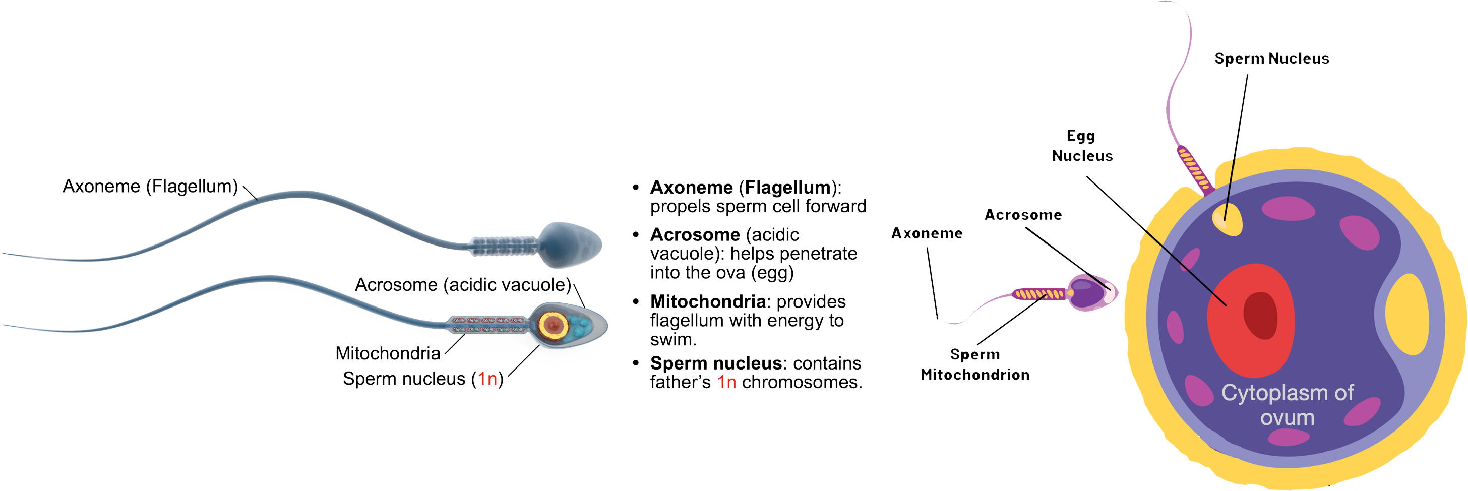
Sperm Cell: The sperm cell is produced in the father’s body and carries 23 chromosomes. It is very small and has a tail called a flagellum, which helps it swim toward the egg cell. Click on the illustration of human sperm below to enlarge the image:
Egg Cell: The egg cell (or ovum) is produced in the mother’s body and has 23 chromosomes. It is much larger than the sperm cell and is filled with nutrients to help the developing embryo grow in its early stages.
The Process of Fertilization
When the sperm cell reaches the egg cell, they combine, this is called fertilization. The sperm’s 23 chromosomes and the egg’s 23 chromosomes join together, making a complete set of 46 chromosomes. This new cell is called a zygote.
The zygote has all the genetic information it needs to develop into a new human. It will continue to divide and grow into an embryo and, eventually, a baby.
Why Is Fertilization Important?
Fertilization is important because it combines genetic material from both parents. This means that the baby will have traits from both the father and the mother, like eye color, hair color, and other characteristics. The combination of chromosomes from both parents also makes every person unique from fertilization onward.
In summary, haploid sperm and egg cells are crucial in fertilization because they combine half of the genetic information from each parent to create a new, complete human organism.
Check Your Understanding: The Menstrual Cycle
- Q: What is the purpose of the menstrual cycle?
 A: It prepares the body for pregnancy by thickening the uterus lining.
- Q: What happens during menstruation?
A: The uterus sheds its lining if no fertilization occurs, resulting in a period.
- Q: When does ovulation usually occur?
A: Around Day 14 of the cycle, when the egg is released from the ovary.
- Q: What happens if an egg is not fertilized?
A: It breaks down, and the menstrual cycle restarts.
- Q: How does the cycle change with age?
A: It starts during puberty (around age 12) and ends with menopause (around age 50).
Check Your Understanding: Fertilization
- Q: What is fertilization?
A: Fertilization is when a sperm cell joins an egg cell, forming a zygote.
- Q: Why do sperm and egg cells each have 23 chromosomes?
A: They are haploid cells, so together they form a complete set of 46 chromosomes.
- Q: Where does fertilization usually occur?
A: In the fallopian tube, after ovulation.
- Q: What is the zygote’s first step after fertilization?
A: It travels to the uterus and starts dividing to form a blastocyst.
- Q: Why is fertilization important for genetic diversity?
A: It combines DNA from both parents, creating a unique individual.
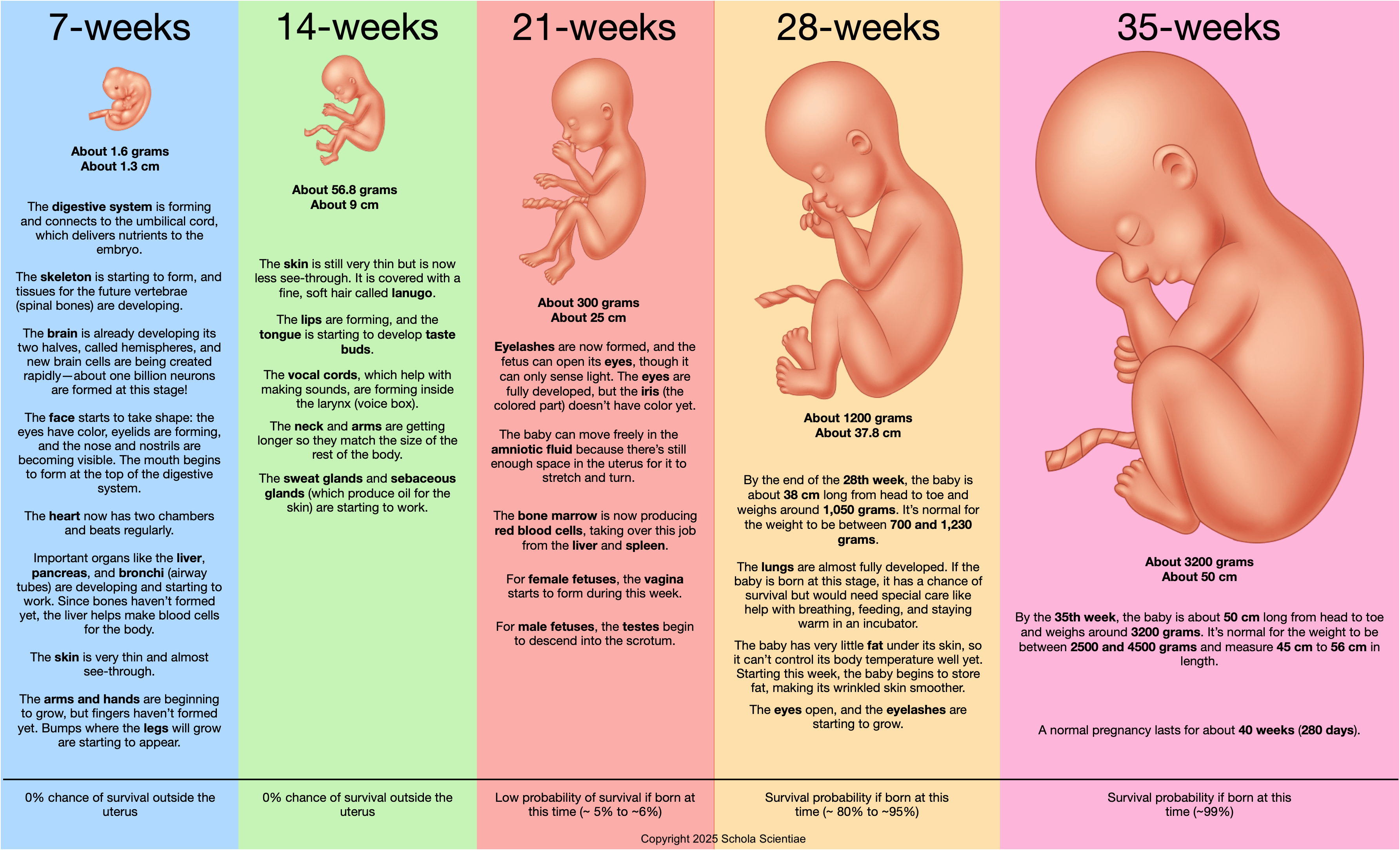
Embryonic Development: First Trimester
Human pregnancy lasts usually lasts approximately nine months and is typically divided into three 3-month intervals referred to as trimesters.
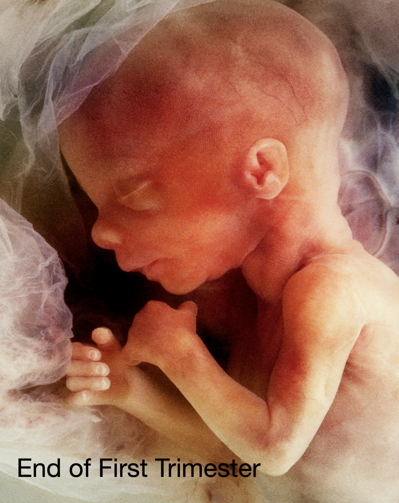
The first trimester is the very beginning of human life, and it covers the first 12 weeks of pregnancy. It’s an incredibly important time because many things happen quickly to help the baby start growing. Let’s break it down step by step.
Week 1-4: The Start
- It all begins when the sperm from the father fertilizes the egg from the mother. This creates a tiny new cell called a zygote.
- The zygote travels down the fallopian tube to the uterus and starts to divide into more and more cells, forming a blastocyst.
- It then implants itself into the thick lining of the uterus, where it will get all the nutrients it needs to grow.
Week 5-8: Big Changes
- During this time, the baby is called an embryo. It’s still tiny, but it’s starting to grow really important parts.
- The heart begins to beat, and blood vessels start to form.
- The brain, spine, and other organs like the liver and lungs are beginning to develop.
- Arm and leg buds appear, which will grow into arms, hands, legs, and feet.
- By the end of this period, the embryo starts to look a little more like a human, though it’s still only about the size of a blueberry!
Week 9-12: The Fetus Stage
- The embryo is now called a fetus. This means it’s still growing but now has most of its important organs starting to work.
- The fingers and toes start to separate and become more defined.
- Bones are beginning to form, and the muscles are developing, which will allow the baby to start moving soon, even if it’s too tiny to feel.
- The baby’s facial features start to become clearer—you can see little ears, eyes, and a nose forming.
- By the end of the first trimester, the baby is about the size of a plum and is growing fast!
Why Is the First Trimester Important?
- It’s when all the major organs and body parts start to form, so it’s a critical time for the baby’s development.
- It’s important for the mother to stay healthy during this time to help the baby grow properly.
In summary, the first trimester is like the building phase, where all the main parts of the baby are starting to form and develop. It’s a very busy time for the tiny new life! Click on the image below to enlarge the embryo (now a fetus) at the end of the first trimester.
 Check Your Understanding
- Q: When does the first trimester occur?
A: It covers the first 12 weeks of pregnancy.
- Q: What major change happens during Weeks 1–4?
A: The zygote forms, becomes a blastocyst, and implants in the uterus.
- Q: When does the heart start beating?
A: The heart begins to beat around Week 5.
- Q: What marks the transition from embryo to fetus?
A: Around Week 9, when most organs start functioning.
- Q: Why is the first trimester important?
A: It’s when all major organs and body systems begin to form.
Fetal Development: Second Trimester
A developing embryo is typically referred to as a fetus after the 10th week of pregnancy. Thus, the entire second and third trimester of pregnancy may be referred to a fetal development.
The second trimester is the period from week 13 to week 26 of pregnancy. This is a time of rapid growth and development for the baby, and it’s often when the mother starts to feel the baby move. Let’s go through what happens step by step!
Weeks 13-16: Growth and Movement
- The baby continues to grow quickly and now looks more like a tiny human. The face becomes more detailed, with eyebrows, eyelashes, and even hair starting to form.
- The baby’s bones are hardening, and the muscles are developing more, allowing the baby to move its arms, legs, and even make little fists.
- By the end of this period, the baby is about the size of an avocado (about 4 to 5 inches long).
 Weeks 17-20: Senses and Reactions
- The baby’s nervous system (brain and nerves) continues to develop, and it starts to react to light and sound.
- The baby’s ears are fully formed, so it can even start hearing sounds like the mother’s heartbeat and voice!
- A layer called vernix caseosa (vernix for short) forms on the baby’s skin to protect it. The skin itself is still very thin.
- At this stage, the baby is around the size of a banana, and the mother may feel kicks and movements for the first time.
Weeks 21-24: Organ Development and Chances of Survival
- The baby’s lungs and digestive system are developing, although they still have more growing to do before they’re ready to work on their own.
- The baby is practicing breathing by inhaling small amounts of amniotic fluid, which helps develop the lungs.
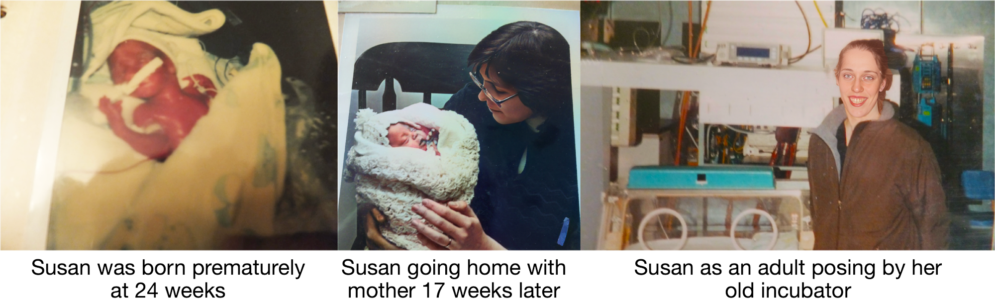
- By week 24, the baby is about the size of an ear of corn. This is also when the baby’s chances of survival if born early, start to improve. Babies born at this stage are called premature, and while they would need special medical help, they have a small chance of survival.
Weeks 25-26: Improving Survival Chances
- During these weeks, the baby’s lungs continue to develop, and the air sacs that are important for breathing start to form. The baby is getting better prepared for life outside the womb.
- The baby also begins to put on more fat, which helps regulate body temperature after birth.
- By the end of the second trimester, the baby is around the size of a cabbage and weighs about 1.5 to 2 pounds.
Survival Chances During the Second Trimester
- Before Week 24: If a baby is born earlier than this, the chances of survival are very low because the lungs and other organs are not developed enough.
- Week 24: Survival rates improve but are still low. With special medical care, a baby born at this stage has a 50% chance of survival.
- Week 26: The survival chances are even better, with some babies having about an 80% chance of surviving if born at this point, though they would still need to be in a neonatal intensive care unit (NICU).
In summary, the second trimester is all about the baby growing bigger, moving more, and developing important organs and systems. As time passes, the baby’s chances of surviving outside the womb improve, especially as it gets closer to the third trimester.
Check Your Understanding
- Q: When does the second trimester occur?

A: From Week 13 to Week 26 of pregnancy.
- Q: What protective layer forms during this stage?
A: The vernix caseosa, a waxy coating that protects the skin.
- Q: When can the baby start hearing sounds?
A: Around Week 18, when the ears are fully formed.
- Q: What is the survival chance if born at Week 24?
A: About 50%, with intensive medical support.
- Q: How does the baby practice breathing?
A: By inhaling small amounts of amniotic fluid.
 Fetal Development: Third Trimester
The third trimester is the final stage of pregnancy, from week 27 to birth, which usually happens around week 40. During this time, the baby’s body matures, gets bigger, and prepares for life outside the womb. Let’s explore what happens in this important stage!
Weeks 27-30: Growth and Brain Development
- The baby continues to grow rapidly, gaining more weight and muscle. At this point, the baby is about the size of a cauliflower and weighs around 2.5 to 3 pounds.
- The brain is developing quickly, becoming more complex and able to control the baby’s body better. The baby’s movements become stronger and more coordinated.
- The baby’s eyes are open, and it can respond to light and sound. It may even turn its head toward bright light coming from outside the mother’s belly.
- The baby’s lungs are still developing but are getting closer to being ready for breathing on their own.
Weeks 31-34: Getting Ready for Birth
- The baby continues to put on more fat, which helps keep it warm after birth. The skin becomes less transparent, and the baby looks more like a newborn.
- The baby’s lungs are almost fully developed, and it’s practicing breathing by taking in amniotic fluid. This practice is important for lung strength.
- The baby’s bones are fully formed but still soft, making it easier for the baby to fit through the birth canal during delivery.
- By this time, the baby’s size is about that of a butternut squash, and it weighs around 4 to 5 pounds.
Weeks 35-37: Maturity and Positioning for Birth
- The baby’s nervous system and lungs continue to mature, and it’s getting ready for life outside the womb.
- Most babies turn into the head-down position, which is the best position for birth.
- The baby’s movements might feel less intense to the mother because it’s running out of space as it grows larger.
- At this point, the baby is about the size of a pineapple and weighs around 5.5 to 6 pounds.
Weeks 38-40: Full Term and Birth
- At week 37, the baby is considered full term, meaning it is fully developed and ready for birth.
- The baby’s organs are mature, especially the lungs, so it can breathe on its own when born.
- The baby is gaining weight rapidly, putting on half a pound or more each week. By birth, most babies weigh between 6 and 9 pounds and are about the size of a watermelon (18-21 inches long).
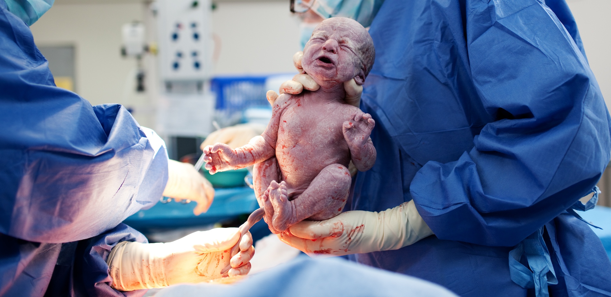
Birth
- When the baby is fully developed and ready, the mother’s body starts a process called labor, where the muscles of the uterus contract to help push the baby out.
- The baby is usually delivered head-first, and after the baby is born, the umbilical cord is cut. This cord was the lifeline that provided the baby with nutrients and oxygen while it was in the womb.
- The baby takes its first breath and begins to cry, which helps clear its lungs and start breathing on its own. The lungs are now fully developed and ready to take in air.
- Click on the image below to enlarge the moment of birth. Notice the thick, creamy vernix. This natural, waxy film protects the baby’s skin from the amniotic fluid during development in the uterus. Notice that the umbilical cord is still attached.
Survival and Health
- Babies born after week 37 have the best chance of being healthy and strong because their organs are fully developed.
- Babies born between week 34 and 37 are often okay but might need some extra help, like staying in a special care nursery.
- Babies born before week 34 are considered premature and may need to spend time in a NICU (Neonatal Intensive Care Unit) to help them grow and develop.
In summary, the third trimester is all about the baby getting bigger, stronger, and fully mature to be ready for birth. It’s the home stretch before the baby enters the world!
Check Your Understanding
- Q: When does the third trimester start?
A: From Week 27 until birth, around Week 40.
- Q: What major brain development happens during this time?
A: The brain grows rapidly, becoming more complex and functional.
- Q: When does the baby turn head-down for birth?
A: Usually between Weeks 35 and 37.
- Q: What is the baby’s status at Week 37?
A: The baby is considered full-term and ready for birth.
- Q: What role does the umbilical cord play?
A: It delivers oxygen and nutrients from the mother to the baby.
Essential Concepts
The Focus Questions in each Investigation are designed to help teachers and students focus on the important concepts. By the end of the CELL, students should be able to answer the following questions:
Modeling the Miricle:
-
Data Interpretation: Based on what you have learned from your reading, class discussions, data tables and graphs, what patterns do you see in fetal mass, length, and organ development between weeks 7, 14, 21, and 28, and what might these patterns tell us about overall fetal growth?
The data shows that as the weeks progress—from week 7 to week 28—the fetus increases significantly in both mass and length, and its organs and limbs develop further. At week 7, the beginnings of organs and limbs are forming, while by week 28, the fetus is much larger and its organs are more fully developed. This pattern indicates that fetal growth accelerates over time, with each developmental stage building on the previous one.
-
Critical Thinking: Why do you think the chances of survival outside the womb increase as the fetus develops, and how do the changes in limb and organ development contribute to this change?
The chances of survival outside the womb increase as the fetus develops because the organs and systems needed for independent life become more mature. For example, as the lungs, heart, and brain develop, the fetus is better prepared to breathe, circulate blood, and regulate its body functions once born. Enhanced limb development also prepares the fetus for movement and coordination, contributing to a higher likelihood of survival.
-
Real-World Application: How can understanding these developmental milestones through data help scientists and healthcare professionals improve prenatal care and support a healthy birth?
By understanding these developmental milestones through data, scientists and healthcare professionals can monitor fetal growth and quickly identify any abnormalities. This helps them provide early interventions or specialized care when needed, ensuring that any issues are addressed promptly to support a healthy birth.
Investigation One:
- Data Interpretation: Examine the karyotype images of a human male and a human female. What differences do you observe in the sex chromosomes, and how do these differences support the concept of genetic sex determination?
When comparing the karyotypes, you’ll notice that both individuals have 22 pairs of autosomes that are identical; however, the female karyotype shows two similar X chromosomes (XX), whereas the male karyotype displays one X chromosome paired with a noticeably smaller Y chromosome (XY). This difference confirms that the presence of the Y chromosome determines male sex.
- Critical Thinking: Considering that the Y chromosome is smaller and contains fewer genes compared to the X chromosome, why do you think this difference exists, and what impact might it have on the expression of genetic traits?
The smaller size of the Y chromosome is due to evolutionary gene loss and specialization—most of its genes are dedicated to triggering male development and sperm production. This means that while the X chromosome carries a wide range of genes affecting many traits, males rely solely on their single X for many functions. Consequently, if a gene on the X is defective, males have no backup copy, which can lead to a higher susceptibility to X-linked genetic disorders.
- Real-World Application: How can understanding karyotypes and the differences between male and female sex chromosomes be applied in medical practice and genetic research?
Knowledge of karyotypes is crucial in diagnosing chromosomal abnormalities, such as Turner syndrome or Klinefelter syndrome, and plays a vital role in genetic counseling. By analyzing the structure and number of chromosomes, healthcare professionals can better assess risks for genetic disorders, tailor treatments, and provide informed advice to patients and their families about potential inherited conditions.
Investigation Two:
- Data Interpretation: Looking at the differences between somatic cells and gametes, what does the process of reduction division reveal about the number of chromosomes in each, and how is this change demonstrated in the lab?”
Reduction division (meiosis) halves the number of chromosomes from 46 in somatic cells (23 pairs) to 23 in gametes, meaning that each gamete contains only one chromosome from each pair. In the lab, this is shown by observing that the gametes have a single set of chromosomes, highlighting how meiosis ensures the correct number of chromosomes is passed on during fertilization.
- Critical Thinking: Why is it crucial for gametes to be haploid rather than diploid, and what potential problems might arise if gametes maintained a diploid number of chromosomes?
It’s crucial for gametes to be haploid because during fertilization, the fusion of two haploid gametes creates a diploid zygote with the proper chromosome number (46 total). If gametes were diploid, fertilization would double the chromosome count, leading to an abnormal number of chromosomes which could result in developmental issues or nonviable embryos.
- Real-World Application: How does understanding the creation of haploid gametes through meiosis help us appreciate the mechanisms behind genetic inheritance and variation in offspring?
By understanding that meiosis produces haploid gametes, we learn how each parent contributes exactly half of the genetic material needed to form a zygote. This process not only ensures that offspring have the correct number of chromosomes but also contributes to genetic variation through mechanisms like crossing over and independent assortment, which are essential for the diversity observed in populations.
Investigation Three:
- Data Interpretation: After observing the fusion of gametes and the subsequent cell divisions, what structural changes do you notice as the zygote transitions into an embryo and later into a fetus?
When examining microscope images, you’ll see that the single-celled zygote begins rapid mitotic divisions to form a cluster of cells that eventually differentiate into various tissues. By the 10th week, these changes lead to recognizable organ structures, and the embryo is then classified as a fetus, which by the 12th week nearly fills the uterus.
- Critical Thinking: Why is the precise process of mitotic cell division after fertilization crucial for proper embryonic development, and what potential issues could arise if these divisions were not accurately regulated?
Accurate mitotic cell divisions ensure that each new cell receives the complete and correct set of genetic instructions necessary for proper differentiation. Disruptions or errors in this process can lead to developmental abnormalities, improper organ formation, or even early miscarriage, highlighting the importance of this tightly controlled process.
- Real-World Application: How does understanding the early stages of human development—from the fusion of gametes to the formation of a fetus—inform practices in prenatal care and developmental diagnostics?
By studying these early developmental stages, healthcare professionals can identify key milestones and potential deviations from normal growth. This knowledge allows for early detection of developmental issues, informs prenatal care strategies, and ultimately aids in improving outcomes for both mothers and babies.
Investigation Four:
- Data Interpretation: Using the life-size anatomical models and ultrasound images provided, what key changes can you identify in fetal organ systems during the second and third trimesters, and how do these changes prepare the fetus for survival after birth?”
During the second and third trimesters, you’ll notice that major organ systems such as the lungs, brain, and heart continue to mature. For example, the lungs develop air sacs for breathing, the brain increases in size and complexity, and the heart grows stronger. These developments, along with an overall increase in size and weight shown in the models, prepare the fetus for the challenges of life outside the womb.
- Critical Thinking: Why might a baby born prematurely at the end of the sixth month have a chance to survive, and which organ systems do you think are most critical for this early viability?
A baby born at the end of the sixth month can survive because key organ systems, especially the lungs, have developed enough to allow breathing, albeit with medical support. Additionally, a developing heart and maturing brain contribute significantly to viability, while modern medical interventions further support these systems until the baby is fully mature.
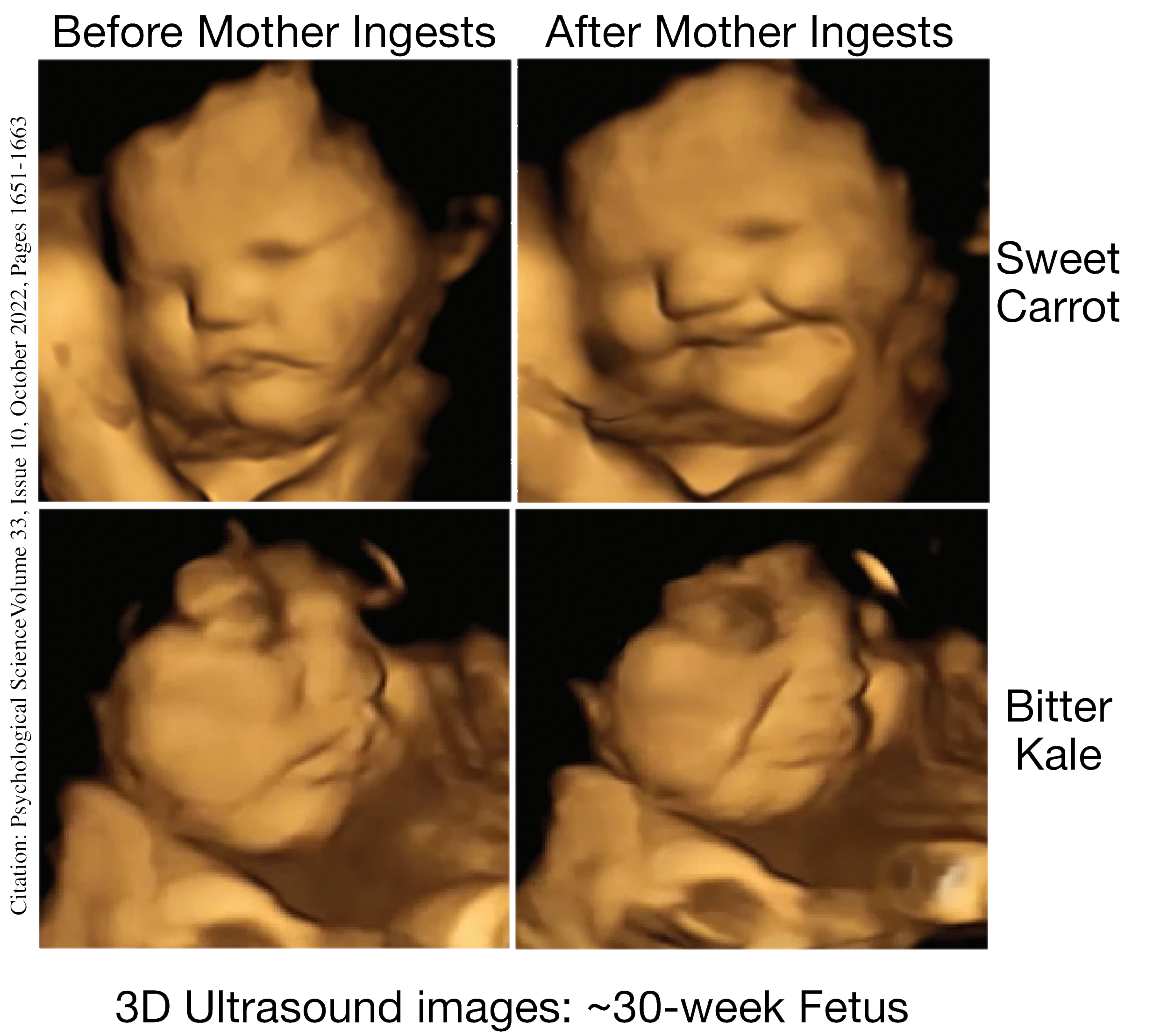
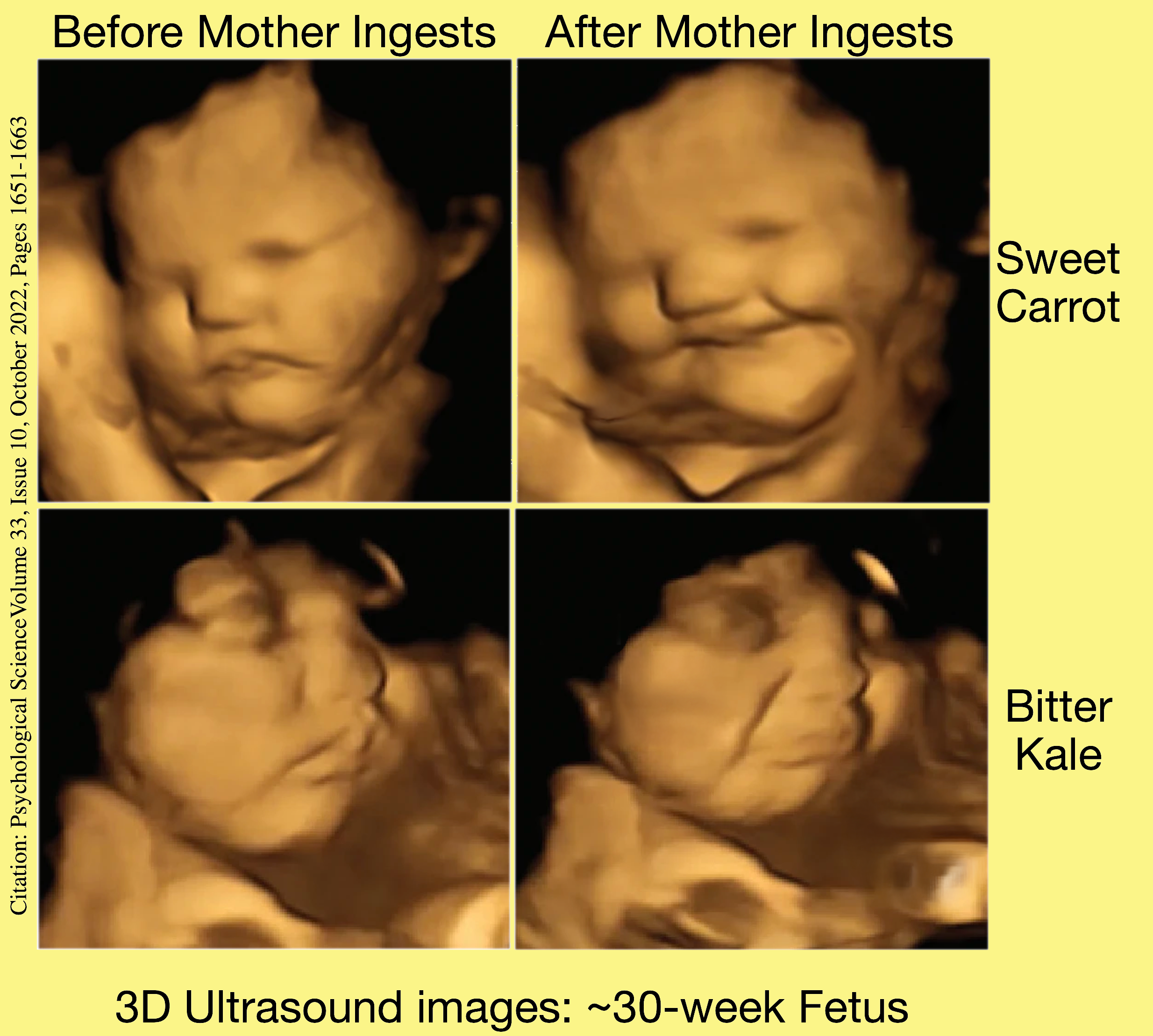
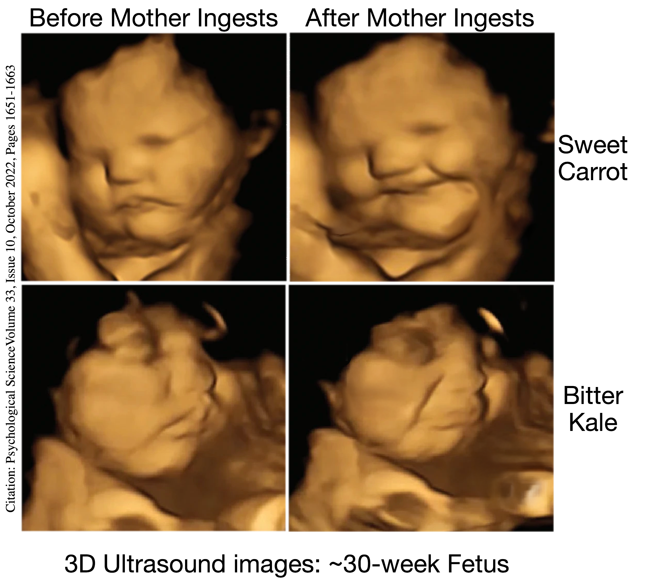
- Real-World Application: How does using ultrasound technology during the later stages of pregnancy benefit both expectant parents and medical professionals in monitoring fetal development?
Ultrasound technology allows doctors to non-invasively track the growth and development of the fetus in real time, identifying any potential issues early. This monitoring provides expectant parents with reassurance and helps medical professionals adjust prenatal care to ensure a safe delivery, illustrating the vital role of technology in modern obstetrics.
CELL Vocabulary
The following list includes key terms that are introduced throughout the CELL. These terms should be used, as appropriate, by teachers and students during everyday classroom discourse.
Amniotic Fluid – Protective liquid surrounding the fetus.
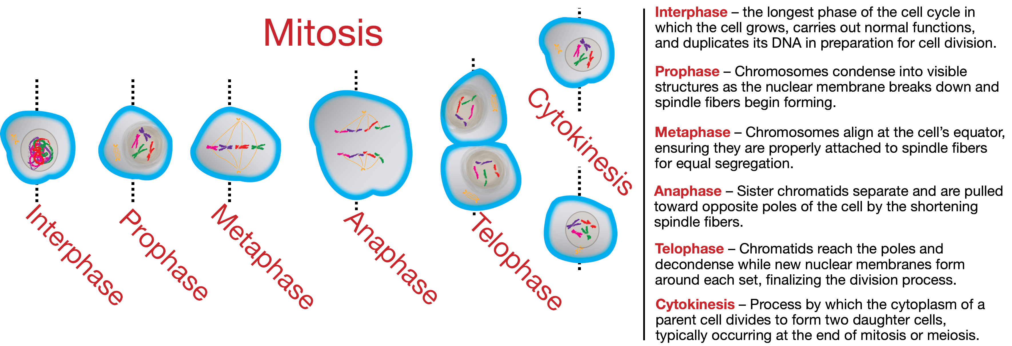
Anaphase – Sister chromatids separate and are pulled toward opposite poles of the cell by the shortening spindle fibers.
Arm and Leg Buds – Early limb structures.
Blastocyst – Cluster of cells formed from the zygote.
Cell Division – Process by which cells reproduce.
Chromosome – DNA structure containing genetic instructions.
Cytokinesis – Process by which the cytoplasm of a parent cell divides to form two daughter cells, typically occurring at the end of mitosis or meiosis.
Daughter Cells – New cells produced after division.
DNA (Deoxyribonucleic Acid) – Molecule carrying genetic material.
Egg Cell (Ovum) – Female reproductive cell, nutrient-rich.
Embryo – Baby’s developmental stage from fertilization to 8 weeks.
Fallopian Tube – Passage for the egg from the ovary to the uterus.
Fertilization – Sperm and egg join to form a zygote.
Fetus – Stage of development from 9 weeks onward.
Full Term – Stage when the baby fully develops (after week 37).
Gametes – Reproductive cells (sperm and egg).
Gene – DNA segment coding for proteins.
Genetic Diversity – Variation resulting from meiosis.
Haploid – Cells with half the usual chromosomes (23 in humans).
Head-Down Position – Optimal fetal position for birth.
Homologous Chromosomes – Paired chromosomes from each parent.
Implantation – Attachment of blastocyst to uterine lining.
Interphase – the longest phase of the cell cycle in which the cell grows, carries out normal functions, and duplicates its DNA in preparation for cell division.
Labor – Uterine contractions for childbirth.
Meiosis – Cell division for reproduction.
Menopause – End of the menstrual cycle, usually at 45–55 years.
Menstrual Cycle – Monthly process preparing for pregnancy.
Menstruation – Shedding of the uterine lining (Days 1–5).
Metaphase – Chromosomes align at the cell’s equator, ensuring they are properly attached to spindle fibers for equal segregation.
Micrograph – Image taken through a microscope.
Mitosis – Cell division for growth and repair.
Neonatal Intensive Care Unit (NICU) – Specialized newborn care unit.
Nervous System – Brain, spinal cord, and nerves. Controls body functions.
Organogenesis – Formation of organs.
Ovulation – Egg release from the ovary (around Day 14).
Pairing – Formation of chromosome pairs during reproduction.
Premature – Baby born before 37 weeks.
Prophase – Chromosomes condense into visible structures as the nuclear membrane breaks down and spindle fibers begin forming.
Puberty – Developmental stage marking reproductive maturity.
Sister Chromatids – Identical copies of chromosomes.
Sperm Cell – Male reproductive cell with a tail (flagellum).
Telophase – Chromatids reach the poles and decondense while new nuclear membranes form around each set, finalizing the division process.
Third Trimester – Final pregnancy stage, week 27 to birth.
Trimester – One of three 3-month pregnancy stages.
Vernix Caseosa – Waxy white coating protecting the baby’s skin.
XX Chromosomes – Female sex chromosomes.
XY Chromosomes – Male sex chromosomes.
Zygote – First cell formed after fertilization.