Teacher Portal
Cellular Organization
Deep Analysis
Deep Analysis
Cellular Organization

Investigation 1
Note: Table A is included for reference.
Table A
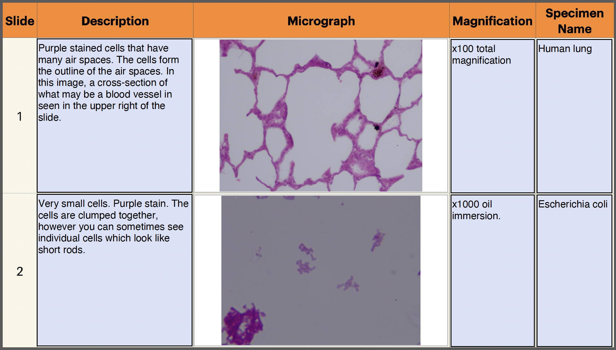
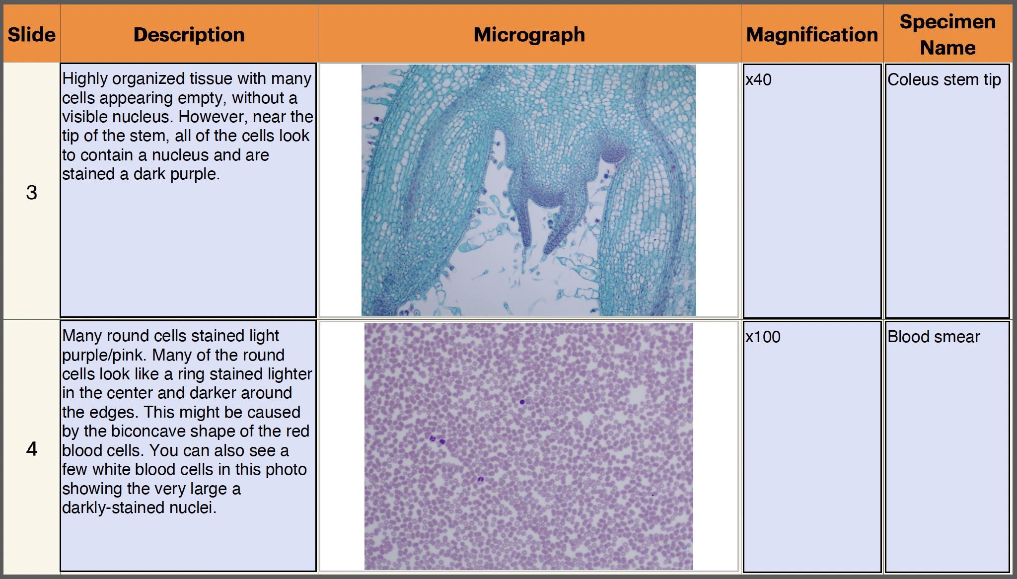
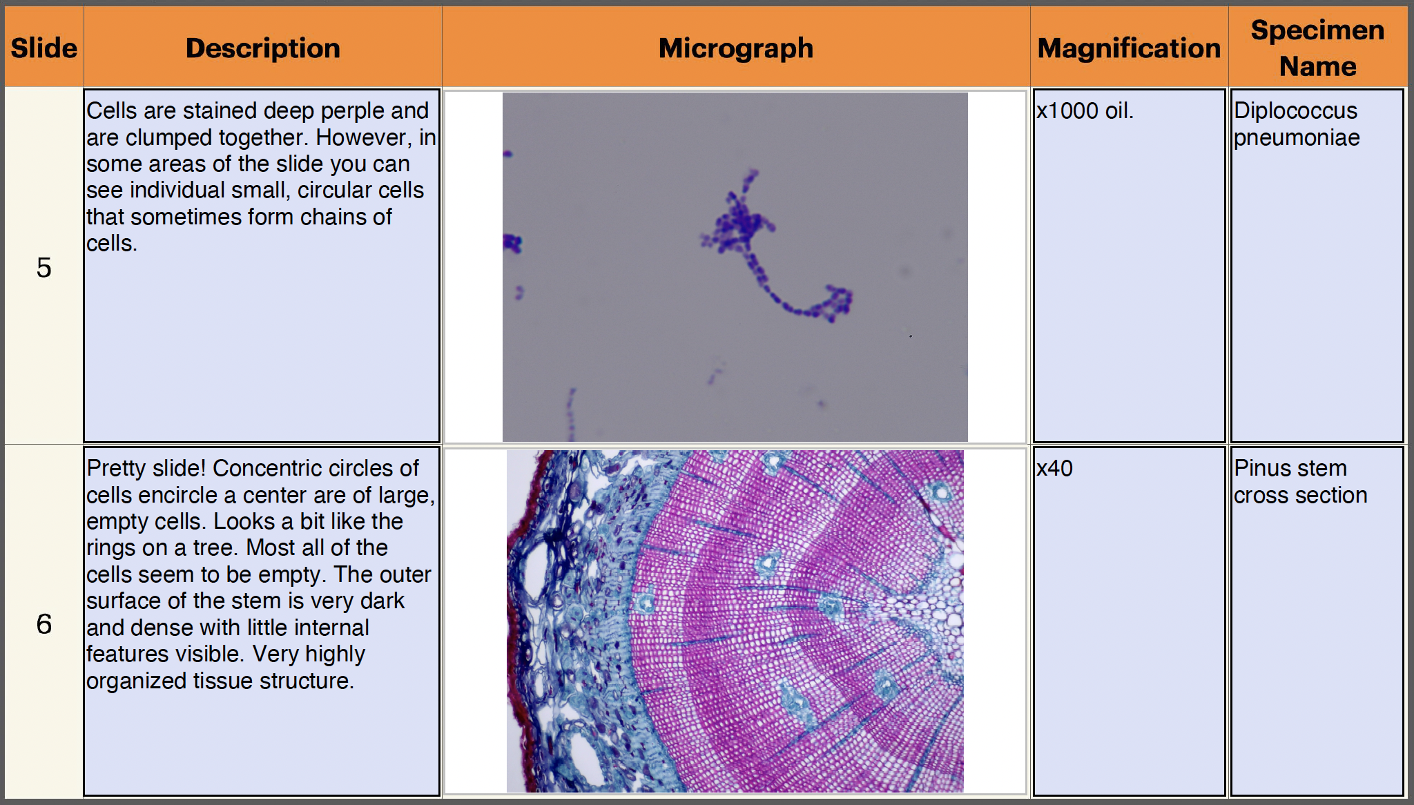
- Compare and contrast the six slides you observed. Student answers will vary. Each of the cells in the six specimens was unique. Upon initial inspection, the cells in slides 2 and 4 appeared similar, because both appeared to consist of many round cells. After observing the slides under 100X, however, one could see that slide 2 consisted of small elliptical cells while slide 4 contained small and large irregularly shaped and round cells with different colored nuclei. The cells in slides 3 and 6 appeared very organized and structured, although the shape of the specimens was quite different.
- Make a prediction. Based on the drawings and descriptions of the slides in Table A, did any of the specimens come from the same organism? Explain your answer. Student answers will vary. Students may conclude that it may be difficult to draw conclusions concerning cells and the organisms they come from simply by observing the specimens under a microscope.
- Was your prediction correct? Student answers will vary.
- What conclusions about cells can you draw after your observations today? Student answers will vary. Students may conclude that cells have different appearances. They are different colors and are organized differently.
- Why did you need to use a microscope? Cells are so small, they are impossible to see without powerful magnification.
- At what magnification were you able to view cells? At a total magnification of 100X, we were able to view cells. Students may need to be reminded of how to calculate total magnification in a microscope. When students look through a microscope, they look through two lenses, one in the eyepiece and one in the objective. In order to calculate total magnification, the magnification of the eyepiece must be multiplied by the magnification of the objective. For example, when using the 10X objective on a microscope with a 10X eyepiece, the total magnification is 100X.
- At what total magnification were you able to view cells with the greatest detail? Our microscopes were able to enlarge the image of the cells to a magnification of 1000X, enabling us to view the cells with the greatest detail.
- How did your view of the cells differ when different objectives were used? Each objective offered a different field of view and resolution. The field of view is the total area observed when viewing the specimen. The resolution is the ability to see fine details in the specimen. As the objectives increased in their refractive power (4X, 10X, 40X, 100X) the field of view decreased, but the resolution of the specimens increased.
- Based on your observations during this investigation, do you feel that you can answer this question? If so, how would you answer? Student answers will vary. Some students may feel that they do not have enough knowledge to determine how cells from one organism differ from those in another. Other students may recall that some cells are a certain color or that some cells are larger than others. Encourage varied responses.
- Based on your observations, do you feel that you can make generalizations about the characteristics of plant cells or of animal cells? For instance, can you determine that all plant cells are a certain size or that all animal cells are arranged in a specific manner? Student answers will vary. Without significant prior knowledge of cells, students may be unable to make generalizations about plant cells and animal cells. However, students may observe that the blood slide contained a non-specific arrangement of cells, whereas the Pinus stem slide contained cells in a specific arrangement. Students also may notice that the arrangement of cells in the Coleus stem slide looked much different from the arrangement of cells in the Pinus stem slide. Students may feel that the Pinus stem slide and the Human lung slide were similar in that they both contained a variety of specifically organized cells.
- What further information or observations do you need to form a scientific conclusion about the differences or similarities between cells of different organisms? What other variables or factors should you investigate? Student answers will vary. Students may feel that they need to look at more slides before they can make reliable conclusions. Students also may feel that they need to learn more about the way slides are prepared to find whether the differences or similarities they observe are due to the properties of the cells or the way the slides were prepared. Students may also wish to research the structures of cells so that they can properly identify and understand the cells they observe.
Investigation 2
Note: Representative micrographs are included for reference. CLICK on any image to enlarge.
As a class, discuss the effect of staining on the characteristics of the specimen.
- Why would it be important for a scientist to know what kind of sectioning was used to prepare a specimen for observation under a microscope? Student answers will vary. Because sectioning can drastically change the appearance of the cells under the microscope, it is important to know what sectioning technique is used. If the type of sectioning is not known or incorrectly identified, it could lead to the misidentification of cells. When comparing cells from different specimens, it would be important to use the same type of sectioning for each so that the differences observed between the cells are actual structural differences within the cell and not the consequence of different sectioning.
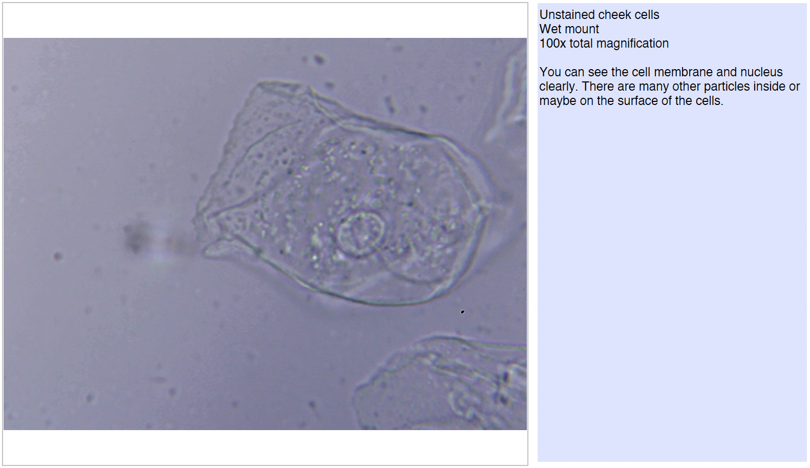
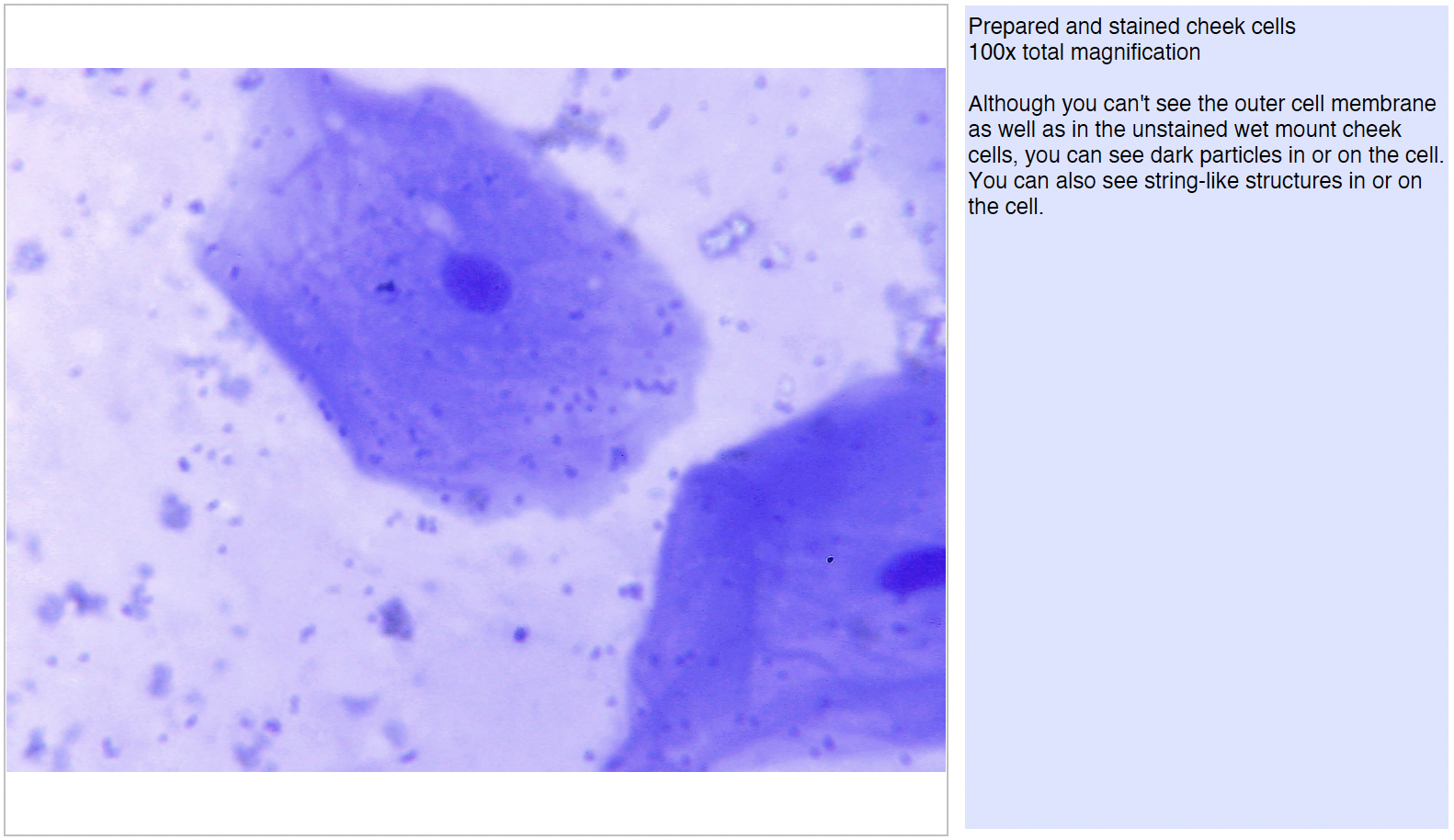
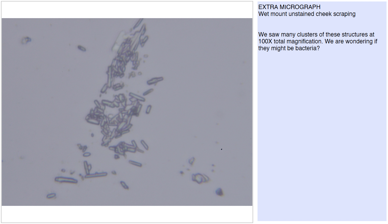
- Did your cheek cells appear different from the prepared cheek cell slide? If so, in what way? Student answers will vary. Students may feel that their cheek cell slide contained fewer cells or that the cells from their slide were a different size than the cells on the prepared slide. Students should notice a distinct difference in the color of the cells. The cells in their own slide were clear and did not contain much color. However, the cells observed on the prepared slide were stained dark pink, with purple nuclei.
- The prepared slide was stained. How did the staining affect your observations of the cheek cells? Student answers will vary. The staining allowed students to observe the cheek cells more easily. The cells were easier to locate on the slide, and students could easily observe the cells’ nuclei on the prepared slide.
- Think back to your observations in Investigation One. Were any of your observations due to the staining of a specific specimen? If students observed color on a slide, their observations were most likely due to the staining of the specimen, rather than the actual color of a cellular structure.
As a class, discuss the effect of sectioning on the characteristics of different specimens.
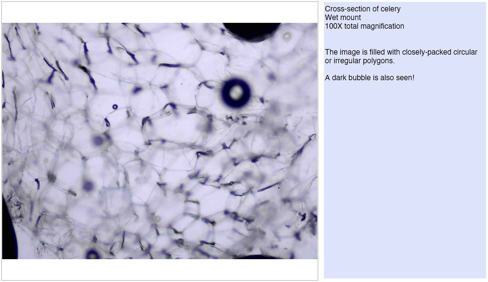
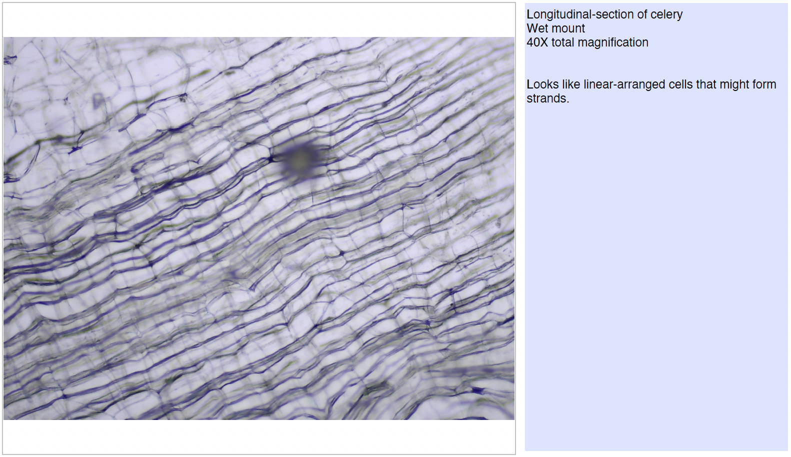
- Did the celery appear different depending on the way it was cut, or sectioned? Yes. When a cross-section of the celery was made, the celery was crescent-shaped. We were able to view small circles scattered across the specimen where the string-like fibers were cut. When a longitudinal section of the celery was made, it was long and rectangular. No small circles were visible. Rather, long stripes stretched across the specimen.
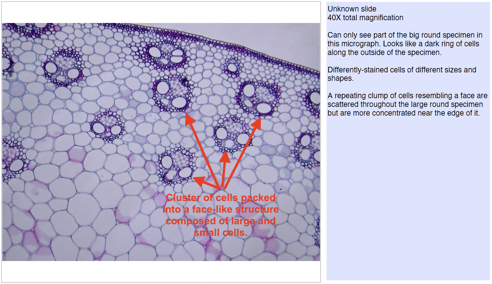
- Did the longitudinal section slide appear different from the cross-section slide of the corn stem? How did this compare to the celery sectioning? Student answers will vary. Just as the celery appeared different depending on the way it was sectioned, so the corn stem appeared differently.
Note: The cross-section of the stem contained many large, somewhat circular cells that appear empty. In various sections of the specimen, students observed clusters of smaller cells and ducts. The longitudinal section of the corn stem contained may long, narrow, rectangular cells which appear empty. In various sections of the specimen, students observed stripes of cells and ducts which are stained purple or red. Although students observed the same structures in each section, the appearance of the structures was changed by the way each specimen was sectioned.
Investigation 3
As a class, discuss the structures that students were able to observe in the six slides they viewed during this investigation.
Note: Micrographs of the six slides are provided below for reference.
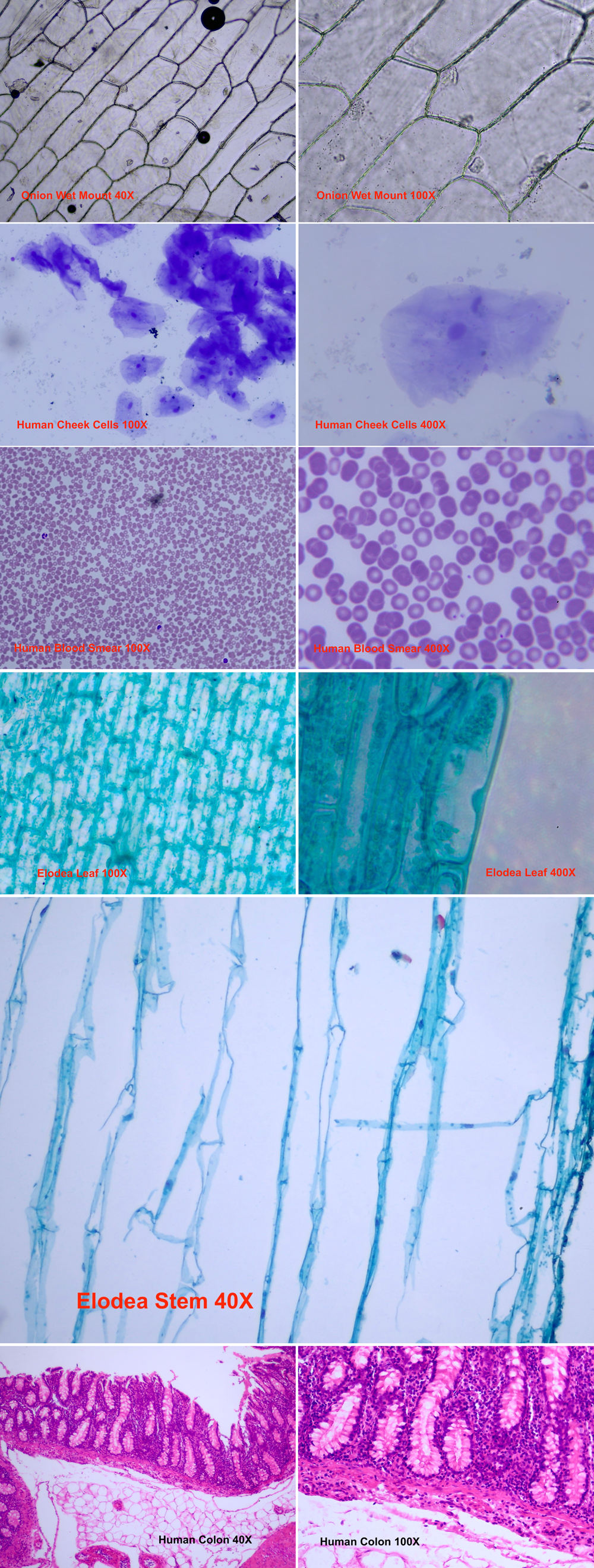
- Based on your knowledge of plant and animal cells, which structures should you have been able to see in all of the plant cells? Were you able to see them? Plant cells generally contain a nucleus, a cell wall, a cell membrane, cytoplasm, chloroplasts, nuclear membrane, centrosome, Golgi, endoplasmic reticulum, central vacuole, microfilaments, microtubules, mitochondria, plasma membrane, and plasmodesmata. However, students were most likely unable to view all of these structures in each of the slides. Visible in the Elodea leaf were the cytoplasm, cell wall, nucleus, cell membrane, and chloroplasts, but it is likely that students would be unable to view a difference between the cell wall and cell membrane. They would, however, be able to observe the cytoplasm and nucleus. The onion did not contain chloroplasts, and students would again be unable to distinguish between the cell wall and the cell membrane. The cytoplasm was more clearly visible in cells that were colored or cells that were stained.
- Why were you unable to distinguish between the cell wall and the cell membrane in the plant cells? Students would have been unable to distinguish between the cell wall and the cell membrane because the space between the two structures is so minimal. Greater magnification or resolution is needed in order to see the separation between the two structures.
- Why were you unable to view the nuclear membrane, centrosome, Golgi, endoplasmic reticulum, central vacuole, microfilaments, microtubules, mitochondria, plasma membrane, and plasmodesmata? Students were unable to distinguish other organelles and structures because they were unstained and smaller in size. Staining and increased resolution would have been needed to observe these structures.
- Why were you unable to view any chloroplasts in the onion cell? To fully answer this question, students must understand the function of chloroplasts and the manner in which onions grow. Chloroplasts are structures responsible for photosynthesis in plants, or food production. Photosynthesis requires light in order to occur. The white onion bulb, or the part of the onion we generally eat, grows underground, with the green leaves growing in the sunlight. Because the white part of the onion does not receive significant sunlight, very little photosynthesis occurs within its cells. The green leaves of the onion contain the cells which are responsible for food production. Therefore, very few chloroplasts are found in the white flesh of the onion.
- Based on your knowledge of plant and animal cells, which structures should you have been able to see in all of the animal cells? Were you able to see them? Animal cells contain cell membranes, nuclei, cytoplasm, endoplasmic reticulum, peroxisomes, centrosomes, mitochondria, lysosomes, microfilaments, microtubules, DNA, nuclear membranes, ribosomes, Golgi apparatus, and plasma membranes. Students were likely able to view some of these structures in all of the specimens, including cell membranes, nuclei, and cytoplasm. Students may have had difficulty seeing the cytoplasm in the cheek cells because the unstained cytoplasm appeared clear. Students also should have noticed that they were able to view nuclei in only some of the blood cells on the blood smear. Inform students that red blood cells do not contain nuclei. The cells with nuclei that students observed were the larger white blood cells rather than the smaller red blood cells. Other organelles and structures may have been difficult to observe because they were unstained or smaller than the resolution of the microscope.
Instruct students to recall one of the questions presented to them at the beginning of Investigation One.
- How are cells from one organism different from or similar to the cells of another? How might you answer the question after you observed the last set of specimens? Based on our observations, all cells had cell membranes, cytoplasm, and nuclei, with the exception of red blood cells, E. coli, and pneumoniae, which had no nuclei. Cells from plants had cell walls and some had observable chloroplasts, but animal cells did not have either of these structures.
As a class, discuss whether or not cells from the same organism are identical.
- Were the two cell samples from the Elodea plant identical? No. The slide of the Elodea leaf was comprised of rectangular cells positioned next to one another. The cells contained cytoplasm and nuclei stained dark blue and purple. The Elodea stem contained somewhat circular cells of various sizes that appeared empty. Students saw different structures and different organizations of cells on each of the two slides, in part because the function of the stem is different than the function of the leaf.
- Compare your observations of the blood smear slide, the cheek cell slide, and the large intestine slide. Were the cells in each of those slides identical? No. The blood cells, cheek cells, and large intestine cells all differed in appearance. The size and shape of the cells varied, as did the structures observed in each cell. The cells in the large intestine, the cheek cells, and the white blood cells contained nuclei, but the red blood cells did not. The red blood cells were disc-shaped and concave, while the cheek cells were irregularly shaped, and the cells in the large intestine were of various shapes.
After students have thought about specific examples, ask the question again.
- Are all cells from the same organism identical? No, the cells in an organism may differ from tissue to tissue.
Ask students to recall all of the cells they viewed throughout Investigations One through Three.
- Why do you think cells from the same organism differ? Cells from the same organism may differ as a result of their function within the organism. For example, red blood cells carry oxygen and carbon dioxide into and away from the tissue. In order to do this, they must be small enough (7 microns) to travel through capillaries. Capillaries can have diameters as small as 5 to 10 microns (μm). Other cells that do not travel through capillaries would not need to be as small.
- How does the arrangement of cells within a tissue affect the function of an organ? Student answers will vary. For example, students may observe differences between the arrangement of cells in the blood smear slide and the large intestine slide. The cells in the blood smear slide seem randomly spread on the slide with little or no organization. However, the cells in the large intestine are very organized in their layout. The organization appears to be specific and the cells create specific structures. These differences may be due to the function of blood and the function of the large intestine. Blood is a liquid that must flow fluidly throughout the body. The seemingly random layout of the cells contributes to this fluidity. The large intestine, however, has the primary function of absorption of water and nutrients. The structures created by the organization of the cells aid in this absorption. Students may attempt to draw conclusions concerning the structure and arrangement of the cheek cells they observed and the function of the cheek tissue. Remind students of the importance of the preparation of the slide when interpreting observations. Given the method used to create the wet mount slide from the cheek cells, it is impossible to know the natural arrangement of the cheek cells within the mouth. In order to draw conclusions about the arrangement and the structure of the cells in the tissue of the mouth, we would need to take a small section of tissue from the cheek and view it, rather than viewing isolated cells.
- Based on your observations, would you agree or disagree with the following statement? Explain your answer. “The function of a tissue or organism depends only on the cellular organelles in the cells.” Student answers will vary. However, students should begin to see that function at the organism or tissue level depends upon both the structures present in individual cells and the organization of cells within a tissue or organ. In addition, students may infer that other factors such as cell-specific genes and proteins also contribute to the specificity of cellular and tissue function.
Investigation 4
- How would you describe the solute contents of the liquids you used to mount the aquatic plant specimens? The liquids each had a different concentration of salt. The tap water contained very little sodium chloride (salt). Although no salt was added to the sample labeled tap water, small amounts of sodium or chloride may have been present initially. As the 2%, 4%, and 8% salt solutions were created, the amount of salt in the water increased. Instruct students to answer question #14 in their Investigation Four Data Record.
- Instruct students to describe what occurred in the aquatic plant leaf when placed in liquids with successively higher concentrations of salt. What did you observe? When observing the aquatic plant leaf in the tap water, students should have been able to identify the cytoplasm, chloroplasts, nuclei and combination cell wall/cell membrane. The chloroplasts were highly visible and contributed the green color to the plant cells and helped to mark the boundary of the cytoplasm and cell membrane. The students observed the cytoplasm with its cellular contents as a small green circular structure within the cell wall. Students may have observed that as the concentration of the salt solution increased, the green structure within each cell wall became smaller. What students observed during this event was the cell membrane and cytoplasm being pulled away from the cell wall as water traveled out of the cell through the cell membrane and the cell wall. As water flowed out of the cell, the turgor pressure within the cytoplasm decreased, the cell membrane was pulled away from the cell wall, and the chloroplasts, cytoplasm, and nucleus were contained within a smaller area of the cell. The boundaries between the cell membrane and cell wall were easily visible as this occurred.
- What conclusions can you draw from your experiments concerning the permeability of the aquatic plant cells to water and sodium chloride? Are the membranes permeable to water? To sodium chloride? The cell membranes were permeable to water. Observations that support this conclusion include the changes in cell (cytoplasm) size as solutions with comparatively higher osmolarities (less water concentration) were added to the aquatic plant specimens. The change in size occurred as water diffused from the cytoplasm through the cell membrane into the external environment. Had the membrane been impermeable to water, students would not have observed a change in the size of the cell membrane and cytoplasm. The cell membranes were impermeable to salt. Had the cell membranes been permeable to salt, salt would have diffused from the external environment into the cell cytoplasms, in an attempt to equalize the osmolarity (the concentration of sodium chloride) on both size the membrane. This would have caused an increase in osmotic pressure inside the cells. Because of the opposing pressure of the cell walls, students would have observed this event as the maintenance of cell membrane and cytoplasmic size. However, the students should indicate that they did not see the maintenance of cytoplasmic size, but rather a decrease. Thus, students should conclude that in the absence of membrane permeability to the sodium chloride, diffusion of water out of the cells was necessary to equalize the osmolarity (sodium chloride concentration) on both sides of the membrane, resulting in a decrease in cytoplasmic size.
- Why did the cytoplasm and cell membranes shrink when the cells were placed in the salt solutions? The cell membranes of the cells in salt solution pulled away from the cell walls as the cells shrank. Because the solutions used to make the slides contained salt, there was a difference in the concentration of salt outside the cell (in the solution) and inside the cell. In other words, the salt solutions contained less water per salt compared to the internal environment of the aquatic plant cells. In order to balance this concentration, water from the cell flowed through the cell membrane and cell wall into the salt solution, causing the salt solution to become more dilute and to be less concentrated.
- Why didn’t the cells shrink as they were placed in the tap water? The cells did not shrink because the difference in concentration of salt was not great enough to cause the movement of water. Because the concentrations outside the cell (in the tap water) and inside the cell were similar, no water movement needed to occur.
- Compare the reaction of the cells to the 2% solution to the reaction to an 8% solution. Explain your observations. When the specimens were placed in the 2% solution, the cell membranes pulled slightly away from the cell walls. When the specimens were placed in an 8% solution, the cell membranes pulled away from the cell walls to a much greater degree. This difference came as a result of the difference in concentrations of salt. The smaller the difference in concentration, the less water that had to move through the cell membrane and cell wall into the surrounding solution.